

International Journal of Clinical Cardiology
An Update on Peripartum Cardiomyopathy in the 21st Century
Kavita Sharma*and Stuart D Russell
Division of Cardiology, Department of Medicine, Johns Hopkins School of Medicine, Johns Hopkins Hospital, USA
*Corresponding author:
Kavita Sharma, MD, Division of Cardiology, Department of Medicine, The Johns Hopkins Hospital, 1800 Orleans Street, Zayed Tower 7125S, Baltimore, MD 21287, USA, Tel: 410-955-5708, Fax: 410-955-3478, E-mail: Ksharma8@jhmi.edu
Int J Clin Cardiol, IJCC-2-034, (Volume 2, Issue 3), Review Article; ISSN: 2378-2951
Received: March 25, 2015 | Accepted: May 30, 2015 | Published: June 02, 2015
Citation: Sharma K, Russell SD (2015) An Update on Peripartum Cardiomyopathy in the 21st Century. Int J Clin Cardiol 2:034. 10.23937/2378-2951/1410034
Copyright: © 2015 Sharma K, et al. This is an open-access article distributed under the terms of the Creative Commons Attribution License, which permits unrestricted use, distribution, and reproduction in any medium, provided the original author and source are credited.
Abstract
Peripartum cardiomyopathy (PPCM) is a pregnancy-associated idiopathic cardiomyopathy secondary to marked left ventricular systolic dysfunction. The disease presents towards the end of pregnancy through the first 5 months post-delivery, with ethnic and geographic variation in incidence and outcomes. While it is relatively rare, PPCM is associated with significant morbidity and can be fatal. The purposes of this review are to summarize the diagnostic criteria, epidemiology, and proposed etiologies of PPCM; present the complications and treatments; and provide a systematic approach to assess risk of PPCM in subsequent pregnancy.
Keywords
Peripartum cardiomyopathy, Pregnancy, Congestive heart failure
Introduction
Heart failure (HF) in the setting of pregnancy has been recognized as far back as the 18thcentury. It was not until 1937, however, that a definitive form of postpartum cardiomyopathy was first described in a case series report by Gouley et al. [1]. The report described 7 women who developed severe, clinical HF in the late months of pregnancy with histologic examination showing widespread and severe focal inflammatory reaction, often with necrosis and followed by fibrosis. In 1971, Demakis et al. described a case series of 27 women with pregnancy-associated cardiomyopathy (PACM) including those who presented with HF in the peripartum period, following which the term "peripartum cardiomyopathy" (PPCM) was first used [2]. The disease remains relatively rare, of unclear etiology, and challenging to recognize and diagnose early. The purpose of this review is to provide an update on the definition and epidemiology of PPCM, proposed mechanisms of disease, clinical manifestations and diagnosis, treatment strategies, and prognosis - particularly with regards to subsequent pregnancies.
Definition
The classic diagnostic criteria for PPCM, defined by Demakis et al., included the following: (1) development of HF in the last month of pregnancy or within 5 months of delivery; (2) absence of an identifiable cause for HF; and (3) absence of recognizable heart disease prior to the last month of pregnancy [3]. In 1997, following a workshop of the National Heart, Lung, and Blood Institute (NHLBI) and Office of Rare Diseases of the National Institutes of Health (NIH), these criteria were expanded to include echocardiography (TTE) criteria of left ventricular systolic dysfunction (left ventricular ejection fraction, LVEF <45%), fractional shortening of <30%, or both [4,5]. The diagnostic criteria for PPCM are summarized in Table 1.
![]()
Table 1: Definition of peripartum cardiomyopathy [3-5]
View Table 1
Although the majority of women with PPCM are diagnosed between the last month of pregnancy and the fifth postpartum month, it is not uncommon for women to present earlier. Elkayam et al. [6] reported that in a series of 123 women with a history of cardiomyopathy diagnosed during pregnancy or in the postpartum period, 100 met criteria for traditional PPCM, while 23 were diagnosed with PACM earlier than the last gestational month, with the earliest diagnosis at weeks. Comparison between the two groups revealed no significant difference in age, ethnic background, obstetric history, or gestational hypertension history. Both groups had similar LVEF at the time of diagnosis, maternal outcome, and recovery rates over time. The early PACM patients did, however, have an increased rate of premature deliveries. This was felt to be related to the earlier development of HF, higher incidence of twin pregnancy, and physician reluctance to continue the pregnancy once the diagnosis was made. Given the otherwise lack of any distinguishing features between the two groups, the authors proposed that perhaps PACM and PPCM in fact represent a spectrum of the same disease [6]. Early presentation of disease has been confirmed in a number of studies and the European Society of Cardiology has expanded their definition of PPCM to include idiopathic cardiomyopathy presenting towards the end of pregnancy and in the months following delivery [7-10].
Epidemiology
The incidence of PPCM in the U.S. is estimated at 1 in 3200 deliveries, with a range from 1 in 1149 to 1 in 4350 live births [11-15]. A study in 2006 reported an increasing incidence in PPCM from 1990 to 2002, likely a reflection of multiple factors including rise in maternal age, rise in multi-gestational pregnancies secondary to infertility treatments, and increased awareness and recognition of the disease itself [13]. In the U.S., the incidence of PPCM has been reported to be up to 16-fold higher in African American women than in non-African American women [14,16]. Higher incidences of PPCM have also been reported in South Africa (up to 1 in 1000) and in Haiti (1 in 3000) [17,18].
Risk factors that are associated with PPCM include advanced maternal age, multiparity, multi-fetal pregnancy, hypertension (chronic, pregnancy induced, or preeclampsia), and African American race [2,6,13,15,19,20]. A recent study of 535 patients with PPCM found that in addition to these established risk factors, substance abuse, asthma, anemia, and auto-immune disease were also associated with PPCM [21].
Etiology
The underlying cause of PPCM remains unknown. A number of proposed mechanisms include: viral myocarditis, abnormal immune response, hemodynamic response to pregnancy, hormonal abnormalities, and malnutrition [4,22-26]. In addition, a large cohort study of PPCM patients has demonstrated elevated plasma levels of tumor necrosis factor-α, Fas-Apo-1, interleukin-6, and C-reactive protein, suggesting an inflammatory component to development of the disease [27].
In 2007 Hilfiker-Kleiner et al. published findings of a novel mechanism for PPCM with a proposal for targeted drug therapy [28]. The investigators demonstrated that female mice with a cardiomyocyte-specific depletion of STAT3 protein developed attenuated expression of manganese sodium dismutase (MnSOD), a known reactive oxygen species (ROS) scavenging enzyme. This resulted in release of cathepsin D (CD), a ubiquitous lysosomal enzyme, which cleaves the hormone prolactin from its 23kDa form to a 16kDa form. This latter form of prolactin induces endothelial cell apoptosis, capillary dissociation, vasoconstriction, and eventually impairs cardiomyocyte metabolism and function, leading to PPCM [28,29]. The group then demonstrated that blockade of prolactin with the dopamine-2D receptor agonist bromocriptine prevented the development of PPCM in mice and subsequently in 6 women at high risk for PPCM due to prior history of PPCM [28]. In 2010 Sliwa et al. published results from a proof-of-concept pilot study in which women diagnosed with PPCM were treated with standard care versus standard care plus bromocriptine [30]. The women who received bromocriptine demonstrated greater recovery of left ventricular ejection fraction (LVEF) compared to the standard therapy group at 6 months. Women in the bromocriptine group experienced far less composite endpoint of poor outcome (death, NYHA function class III/IV, or LVEF <35%) at 6 months compared to the standard therapy group. The findings warrant further study in a large, randomized control trial.
Genetics
PPCM was previously classified as a non-genetic form of dilated cardiomyopathy (DCM). Recent studies, however, have shown familial clustering of PPCM [9,31]. In a study of 4110 women from 520 families of patients with DCM, 45 were identified as having either PACM or PPCM, and familial clustering was found in 23 (55%) of the 42 unrelated cases [32]. Resequencing data was available for 19 women from this group, of which 6 had mutations identified in genes that have been shown to be associated with DCM. Subsequent studies have further supported these findings and additionally shown that screening of first degree relatives of patients with PPCM has resulted in unmasking familial DCM [9,33]. These studies would suggest that there are likely genetic factors implicit in the development of PPCM in a proportion of these patients.
Clinical Presentation and Diagnosis
Most patients diagnosed with PPCM present with typical signs and symptoms of HF. These symptoms often overlap with those of pregnancy itself, which can result in a delay or missed diagnosis and ultimately the development of PPCM-associated complications [15]. Early presentation of PPCM during pregnancy is being increasingly recognized as demonstrated by Elkayam et al., with no significant differences in the clinical characteristics or outcomes between those who present within the traditional PPCM timeframe and those who present earlier [6].
As with any new presentation of HF, the standard evaluation for any woman suspected of PPCM includes a detailed history and physical examination, 12-lead electrocardiogram, 2-dimensional TTE with Doppler, chest radiograph, and the following laboratory tests at baseline: complete blood count, urinalysis, serum electrolytes (including calcium and magnesium), blood urea nitrogen, serum creatinine, blood glucose, liver function tests, lipid profile, and thyroid-stimulating hormone [34].
From a diagnostic testing standpoint, the electrocardiogram in PPCM patients typically shows sinus tachycardia with non-specific ST-segment and T-wave changes [11]. A TTE study must show depressed LVEF for diagnosis. Other TTE findings may include dilation of the LV and/or other chambers, valvular compromise including moderate to severe mitral and tricuspid valve regurgitation, mild to moderate pulmonic valve regurgitation, and pulmonary hypertension [5,10]. Chest radiograph will often show cardiomegaly, pulmonary venous congestion, and occasionally pulmonary edema or pleural effusion [11].
Further testing should be obtained based on the clinical presentation and risks of the individual patient. Both endomyocardial biopsy and coronary angiography should not be considered parts of the routine evaluation of patients with suspected PPCM [34]. The ACC/AHA guidelines currently recommend endomyocardial biopsy only in clinical scenarios where determining a specific etiology for HF may have treatment implications. This includes the setting of unexplained, new-onset heart failure of less than 2 weeks duration associated with a normal-sized or dilated left ventricle in addition to hemodynamic compromise, and new-onset heart failure of 2 weeks to 3 months duration associated with a dilated left ventricle and new ventricular arrhythmias, second- or third-degree heart block, or failure to respond to usual care within 1 to 2 weeks [35]. Reports of endomyocardial biopsy findings in women with PPCM have demonstrated variable rates of myocarditis. In a series of 57 endomyocardial biopsy samples evaluated for suspected PPCM, Rizeq et al. report 34 patients were confirmed to have PPCM with 3 (8.8%) biopsy samples demonstrating focal myocarditis [8]. There were no significant morphological differences in degrees of myocyte hypertrophy, fibrosis, coronary artery pathology, or small intramyocardial vessels between samples from women with PPCM and those with idiopathic dilated cardiomyopathy. Coronary angiography is only recommended as part of the evaluation for new onset HF if there is a strong clinical suspicion of coronary artery disease and therefore should not be considered part of the routine evaluation of patients with suspected PPCM [34]. There is limited data on the use of cardiac magnetic resonance imaging (cMRI) in the diagnosis of PPCM, although findings of myocardial delayed enhancement on cMRI have been reported and cMRI may be used to identify thrombus in the setting of left ventricular dysfunction in PPCM [36]. The use of gadolinium is not recommended as it can cross the placenta [37].
From the standpoint of serum biomarkers, B-type natriuretic peptide (BNP) levels remain unchanged for the most part during normal pregnancy, can be slightly elevated in the setting of preeclampsia, and become markedly elevated in PPCM, as is the case in other forms of HF [38]. Troponin levels may be elevated in PPCM, depending on the degree of myocardial injury incurred at the time of diagnosis [39]. Of most importance from a diagnostic standpoint is that a high index of suspicion for heart failure be maintained at the first signs and symptoms of HF. Common symptoms such as dyspnea, fatigue, and mild edema should not be assumed to be related to pregnancy itself, lack of sleep, or other conditions such as bronchitis; PPCM must also be considered in this patient population.
Complications and Prognosis
PPCM is a significant cause of morbidity and mortality in pregnant patients. Complications associated with the disease include severe HF, cardiogenic shock, arrhythmias, thromboembolic complications, cardiopulmonary arrest, and death [20,40]. Kao et al. recently reported that compared to non-PPCM patients, those with PPCM have higher rates of stillbirth, Cesarean section, major adverse events, and longer length of hospital stay [21]. In a series of 182 patients with PPCM, the rate of major adverse events was reported to be 25%, with 80% of these events occurring in the first six months after the diagnosis [40]. Mortality rates reported to date are around 15%, which is less than that associated with other forms of cardiomyopathy, [14,41]. In a series of 17 cases of death in PPCM patients, 18% of deaths occurred within one week of delivery and 87% within 6 months [42]. The cause of death in these cases was either sudden cardiac death or progressive HF. Geographical differences exist with higher mortality rates reported in Haiti, South Africa, and Turkey compared with the U.S. [21]. Mortality risk is increased with older age, multiparity, African American race, severe LV dysfunction (LVEF <25%), and delay in diagnosis [42].
The rate of cardiac transplantation in PPCM patients is anywhere from 6-23% [20,43]. In a recent report from the United Network for Organ Sharing (UNOS) database, Rasmussen et al. report that patients with PPCM who underwent cardiac transplant were younger, had higher sensitization, required more intense cardiovascular support pre-transplant, and had higher listing status [44]. PPCM patients had more post-transplant rejection (during index hospitalization and in the year to follow), inferior graft survival, and worse age-adjusted overall survival.
With regards to recovery of LV function, recent publications indicate that at least 50% of PPCM patients have improvement in cardiac function within 2 to 6 months following the diagnosis [6,20]. Amos et al. reported outcomes a single-center experience on 55 PPCM patients and found that factors associated with lack of LV function recovery were LVEF at 2 months, LV end-diastolic dimension >5.6cm, the presence of LV thrombus, and African-American race. LVEF at the time of diagnosis was not predictive of recovery [20].
Subsequent Pregnancies
Given that PPCM is a relatively rare form of HF, information on the outcome of subsequent pregnancies is limited. In 2001, Elkayam at al. reported the outcomes of 44 women who had PPCM with a total of 60 subsequent pregnancies [45]. The women were divided into two groups: group 1 consisted of those in whom LV function had returned to normal; group 2 consisted of those with persistent LV dysfunction. Both groups were noted to have a reduction in mean LVEF measurement with the subsequent pregnancy, however, a substantial reduction in LVEF (>20%) was seen in 21% of group 1 and 44% of group 2. A higher proportion of patients in group 2 experienced symptoms of HF. Mortality rate for group 1 was 0%, while for group 2 was 19%. Subsequent studies have shown worsening of HF symptoms in nearly 30% of women with history of PPCM during subsequent pregnancies [43,46]. A recent study by Fett et al. of 61 subsequent pregnancies in women with prior PPCM reported a relapse rate of 29% with a significantly higher rate in women with LVEF <55% [47]. It has been suggested that demonstration of normal contractile reserve in those patients with recovered LV function may portend a better outcome; however this has not been validated in a study to date. The findings of the aforementioned studies suggest that subsequent pregnancies are associated with a risk for recurrent, and/or persistent HF symptoms and LV dysfunction, and even death.
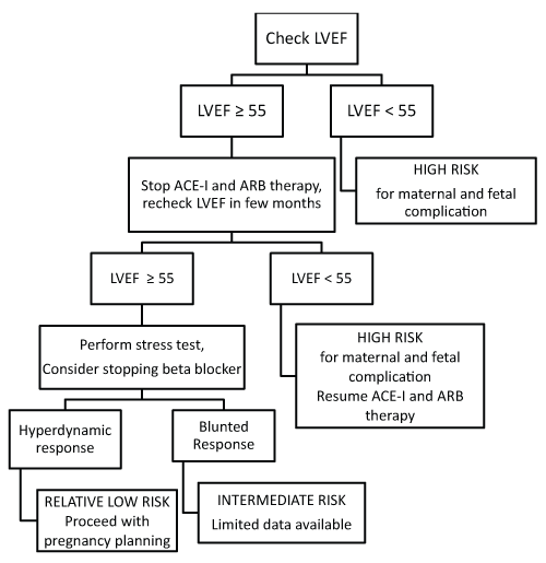
A systematic approach to patients who wish to become pregnant again is depicted in Figure 1. In summary, a baseline echocardiogram should be obtained, along with a serum BNP level. Women with preserved LV function (>55%) should have a repeat echocardiogram a few months after discontinuing angiotensin-converting enzyme (ACE) inhibitors or angiotensin receptor blockers (ARBs), as these medications are contraindicated in pregnancy. If at that time LVEF is still preserved, consideration should be given to stopping beta blocker therapy and performing a stress echocardiogram to assess contractile reserve. Should the findings of a stress test demonstrate hyperdynamic response, the patient would be considered relatively low risk for maternal and fetal adverse outcomes and may be counseled to proceed with pregnancy planning with close monitoring and follow up. They should be counseled that they still have a risk of recurrence but that they are relatively lower risk. Should the response to stress test be no augmentation of LVEF, or a blunted response, then the patient would be considered moderate risk, a group for which we have limited to no data to guide recommendations. If the baseline LVEF is <55% at the start of subsequent pregnancy planning, the patient would be considered high risk and should be strongly advised against second pregnancy, or should be counseled accordingly regarding options including termination of pregnancy. In all risk groups, any subsequent pregnancy warrants follow up echocardiography at a minimum during the early second and third trimesters, the last gestational month, and for any development or progression in HF symptoms[15]. Repeat BNP may be helpful in distinguishing HF symptoms from pregnancy symptoms.
.
Figure 1: Algorithm for considering pregnancy in women who have recovered from their initial cardiomyopathy
View Figure 1
Treatment
Treatment for PPCM is based on guidelines for standard HF treatment, including ACE-inhibitors, ARBs, beta-blockers, spironolactone, digoxin, diuretics, vasodilators and inotropes if needed [48]. Initiation and treatment with any HF therapies should be done under the supervision of a multi-disciplinary team including the patient's cardiologist and obstetrician. The mainstays of treatment for acute decompensated PPCM include diuretics and renin-angiotensin inhibiting agents for patients that are hemodynamically stable. For patients in cardiogenic shock inotropes with or without mechanical support may be required. Once stabilized, beta-blockers, aldosterone-antagonists and additional therapies should be initiated and up-titrated as tolerated in the outpatient setting, particularly in the setting of persistent LV dysfunction. HF drug therapies and their pregnancy and lactation safety profiles are summarized in Table 2.
![]()
Table 2: Heart failure drug safety profile in pregnancy and lactation
View Table 2
A small study of 11 PPCM patients demonstrated that IV immune globulin resulted in improvement in LVEF in the treatment group compared to standard therapy [49]. Another small study has reported improved outcomes in the combined endpoint of persistent LV dysfunction, NYHA functional class, and death with pentoxifylline therapy for PPCM [50]. Sliwa et al. reported the findings of treatment with bromocriptine in a proof-of-concept study, discussed earlier in this review [30]. Larger clinical studies are warranted to clearly establish the role of these newer therapies for PPCM.
As PPCM is associated with a risk of arrhythmias and sudden cardiac death, current recommendations are to consider implantation of an implantable cardioverter-defibrillator (ICD) in patients with persistent LV dysfunction despite optimal HF therapy [40,42,51]. While LV recovery occurs in most patients between 2 to 6 months postpartum, an individual's likelihood of recovery is still relatively unpredicatable [20]. Therefore, it is reasonable to consider a wearable external defibrillator or a subcutaneous ICD as a bridge to recovery or ICD implantation, should LV dysfunction persist.
There are reports of an increased incidence of thrombus formation in patients who develop PPCM and thus consideration of anticoagulation is warranted [1,2,48,49]. For patients who develop fulminant HF symptoms and hemodynamic instability during or after pregnancy, therapies such as intra-aortic balloon pump, LV assist devices, and extracorporeal membrane oxygenation may be considered for use as a bridge to recovery or heart transplant. Outcomes post heart transplant have been discussed earlier in this review.
Conclusions
PPCM is a rare but potentially lethal disease that remains a challenge to diagnose, prognosticate, and treat. The latest updates to the definition of the disease itself now broaden the timeframe of diagnosis to include earlier stages of pregnancy. It is increasingly recognized that the condition is often diagnosed late which may portend a poor prognosis. While the etiology of the disease remains unclear, a novel mechanism involving a mutation in a protein implicated in cleaving prolactin resulting in myocardial damage warrants further study. In addition, a genetic component is likely present, with familial clustering of PPCM reported in families with DCM. Predictors of recovery of LV function include race, LV function, and LV dimensions at follow up. The mainstay of treatment today include standard HF therapies, however, newer therapies such as bromocriptine, IV immunoglobulin, and pentoxifylline are on the horizon and warrant further study. We suggest a large, international registry be created to follow PPCM patients longitudinally. At present, the NHLBI sponsors the Peripartum Cardiomyopathy Network (PCN), a network of physicians and nurses at clinical sites across the United States and Canada dedicated to both clinical care and investigation of PPCM. Such a database will be beneficial in providing baseline characteristics of these patients, family history that would allow for genetic testing in the future and post-partum assessments including echocardiography and/or stress test data to help better predict morbidity and mortality in this population. Lastly, a registry would eventually serve as a resource for the conduction of clinical treatment trials, an area where there is much need in this high risk population.
References
-
Gouley BA, McMillan TM, Bellet S (1937) Idiopathic myocardial degeneration associated with pregnancy and especially the puerperium. Am J Med Sci 194: 185-199.
-
Demakis JG, Rahimtoola SH, Sutton GC, Meadows WR, Szanto PB, et al. (1971) Natural course of peripartum cardiomyopathy. Circulation 44: 1053-1061.
-
Demakis JG, Rahimtoola SH (1971) Peripartum cardiomyopathy. Circulation 44: 964-968.
-
Pearson GD, Veille JC, Rahimtoola S, Hsia J, Oakley CM, et al. (2000) Peripartum cardiomyopathy: National Heart, Lung, and Blood Institute and Office of Rare Diseases (National Institutes of Health) workshop recommendations and review. JAMA 283: 1183-1188.
-
Hibbard JU, Lindheimer M, Lang RM (1999) A modified definition for peripartum cardiomyopathy and prognosis based on echocardiography. Obstet Gynecol 94: 311-316.
-
Elkayam U, Akhter MW, Singh H, Khan S, Bitar F, et al. (2005) Pregnancy-associated cardiomyopathy: clinical characteristics and a comparison between early and late presentation. Circulation 111: 2050-2055.
-
Forssell G, Laska J, Olofsson C, Olsson M, Mogensen L (1994) Peripartum cardiomyopathy--three cases. J Intern Med 235: 493-496.
-
Rizeq MN, Rickenbacher PR, Fowler MB, Billingham ME (1994) Incidence of myocarditis in peripartum cardiomyopathy. Am J Cardiol 74: 474-477.
-
van Spaendonck-Zwarts KY, van Tintelen JP, van Veldhuisen DJ, van der Werf R, Jongbloed JD, et al. (2010) Peripartum cardiomyopathy as a part of familial dilated cardiomyopathy. Circulation 121: 2169-2175.
-
Sliwa K, Hilfiker-Kleiner D, Petrie MC, Mebazaa A, Pieske B, et al. (2010) Current state of knowledge on aetiology, diagnosis, management, and therapy of peripartum cardiomyopathy: a position statement from the Heart Failure Association of the European Society of Cardiology Working Group on peripartum cardiomyopathy. Eur J Heart Fail 12: 767-778.
-
Witlin AG, Mabie WC, Sibai BM (1997) Peripartum cardiomyopathy: an ominous diagnosis. Am J Obstet Gynecol 176: 182-188.
-
Chapa JB, Heiberger HB, Weinert L, Decara J, Lang RM, et al. (2005) Prognostic value of echocardiography in peripartum cardiomyopathy. Obstet Gynecol 105: 1303-1308.
-
Mielniczuk LM, Williams K, Davis DR, Tang AS, Lemery R, et al. (2006) Frequency of peripartum cardiomyopathy. Am J Cardiol 97: 1765-1768.
-
Brar SS, Khan SS, Sandhu GK, Jorgensen MB, Parikh N, et al. (2007) Incidence, mortality, and racial differences in peripartum cardiomyopathy. Am J Cardiol 100: 302-304.
-
Elkayam U (2011) Clinical characteristics of peripartum cardiomyopathy in the United States: diagnosis, prognosis, and management. J Am Coll Cardiol 58: 659-670.
-
Gentry MB, Dias JK, Luis A, Patel R, Thornton J, et al. (2010) African-American women have a higher risk for developing peripartum cardiomyopathy. J Am Coll Cardiol 55: 654-659.
-
Desai D, Moodley J, Naidoo D (1995) Peripartum cardiomyopathy: experiences at King Edward VIII Hospital, Durban, South Africa and a review of the literature. Trop Doct 25: 118-123.
-
Fett JD, Christie LG, Carraway RD, Murphy JG (2005) Five-year prospective study of the incidence and prognosis of peripartum cardiomyopathy at a single institution. Mayo Clin Proc 80: 1602-1606.
-
Sliwa K, Fett J, Elkayam U (2006) Peripartum cardiomyopathy. Lancet 368: 687-693.
-
Amos AM, Jaber WA, Russell SD (2006) Improved outcomes in peripartum cardiomyopathy with contemporary. Am Heart J 152: 509-513.
-
Kao DP, Hsich E, Lindenfeld J (2013) Characteristics, adverse events, and racial differences among delivering mothers with peripartum cardiomyopathy. JACC Heart Fail 1: 409-416.
-
Geva T, Mauer MB, Striker L, Kirshon B, Pivarnik JM (1997) Effects of physiologic load of pregnancy on left ventricular contractility and remodeling. Am Heart J 133: 53-59.
-
Mone SM, Sanders SP, Colan SD (1996) Control mechanisms for physiological hypertrophy of pregnancy. Circulation 94: 667-672.
-
Ansari AA, Neckelmann N, Wang YC, Gravanis MB, Sell KW, et al. (1993) Immunologic dialogue between cardiac myocytes, endothelial cells, and mononuclear cells. Clin Immunol Immunopathol 68: 208-214.
-
Melvin KR, Richardson PJ, Olsen EG, Daly K, Jackson G (1982) Peripartum cardiomyopathy due to myocarditis. N Engl J Med 307: 731-734.
-
Midei MG, DeMent SH, Feldman AM, Hutchins GM, Baughman KL (1990) Peripartum myocarditis and cardiomyopathy. Circulation 81: 922-928.
-
Sliwa K, F�rster O, Libhaber E, Fett JD, Sundstrom JB, et al. (2006) Peripartum cardiomyopathy: inflammatory markers as predictors of outcome in 100 prospectively studied patients. Eur Heart J 27: 441-446.
-
Hilfiker-Kleiner D, Kaminski K, Podewski E, Bonda T, Schaefer A, et al. (2007) A cathepsin D-cleaved 16 kDa form of prolactin mediates postpartum cardiomyopathy. Cell 128: 589-600.
-
Hilfiker-Kleiner D, Sliwa K, Drexler H (2008) Peripartum cardiomyopathy: recent insights in its pathophysiology. Trends Cardiovasc Med 18: 173-179.
-
Sliwa K, Blauwet L, Tibazarwa K, Libhaber E, Smedema JP, et al. (2010) Evaluation of bromocriptine in the treatment of acute severe peripartum cardiomyopathy: a proof-of-concept pilot study. Circulation 121: 1465-1473.
-
Pearl W (1995) Familial occurrence of peripartum cardiomyopathy. Am Heart J 129: 421-422.
-
Morales A, Painter T, Li R, Siegfried JD, Li D, et al. (2010) Rare variant mutations in pregnancy-associated or peripartum cardiomyopathy. Circulation 121: 2176-2182.
-
Anderson JL, Horne BD (2010) Birthing the genetics of peripartum cardiomyopathy. Circulation 121: 2157-2159.
-
Hunt SA, Abraham WT, Chin MH, Feldman AM, Francis GS, et al. (2005) ACC/AHA 2005 Guideline Update for the Diagnosis and Management of Chronic Heart Failure in the Adult: a report of the American College of Cardiology/American Heart Association Task Force on Practice Guidelines (Writing Committee to Update the 2001 Guidelines for the Evaluation and Management of Heart Failure): developed in collaboration with the American College of Chest Physicians and the International Society for Heart and Lung Transplantation: endorsed by the Heart Rhythm Society. Circulation 112: e154-235.
-
Cooper LT, Baughman KL, Feldman AM, Frustaci A, Jessup M, et al. (2007) The role of endomyocardial biopsy in the management of cardiovascular disease: a scientific statement from the American Heart Association, the American College of Cardiology, and the European Society of Cardiology. Circulation 116: 2216-2233.
-
Marmursztejn J, Vignaux O, Goffinet F, Cabanes L, Duboc D (2009) Delayed-enhanced cardiac magnetic resonance imaging features in peripartum cardiomyopathy. Int J Cardiol 137: e63-64.
-
Chen MM, Coakley FV, Kaimal A, Laros RK Jr (2008) Guidelines for computed tomography and magnetic resonance imaging use during pregnancy and lactation. Obstet Gynecol 112: 333-340.
-
Hameed AB, Chan K, Ghamsary M, Elkayam U (2009) Longitudinal changes in the B-type natriuretic peptide levels in normal pregnancy and postpartum. Clin Cardiol 32: E60-62.
-
Hu CL, Li YB, Zou YG, Zhang JM, Chen JB, et al. (2007) Troponin T measurement can predict persistent left ventricular dysfunction in peripartum cardiomyopathy. Heart 93: 488-490.
-
Goland S, Modi K, Bitar F, Janmohamed M, Mirocha JM, et al. (2009) Clinical profile and predictors of complications in peripartum cardiomyopathy. J Card Fail 15: 645-650.
-
Felker GM, Thompson RE, Hare JM, Hruban RH, Clemetson DE, et al. (2000) Underlying causes and long-term survival in patients with initially unexplained cardiomyopathy. N Engl J Med 342: 1077-1084.
-
Whitehead SJ, Berg CJ, Chang J (2003) Pregnancy-related mortality due to cardiomyopathy: United States, 1991-1997. Obstet Gynecol 102: 1326-1331.
-
Habli M, O'Brien T, Nowack E, Khoury S, Barton JR, et al. (2008) Peripartum cardiomyopathy: prognostic factors for long-term maternal outcome. Am J Obstet Gynecol 199: 415.
-
Rasmusson K, Brunisholz K, Budge D, Horne BD, Alharethi R, et al. (2012) Peripartum cardiomyopathy: post-transplant outcomes from the United Network for Organ Sharing Database. J Heart Lung Transplant 31: 180-186.
-
Elkayam U, Tummala PP, Rao K, Akhter MW, Karaalp IS, et al. (2001) Maternal and fetal outcomes of subsequent pregnancies in women with peripartum cardiomyopathy. N Engl J Med 344: 1567-1571.
-
Modi KA, Illum S, Jariatul K, Caldito G, Reddy PC (2009) Poor outcome of indigent patients with peripartum cardiomyopathy in the United States. Am J Obstet Gynecol 201: 171.
-
Fett JD, Fristoe KL, Welsh SN (2010) Risk of heart failure relapse in subsequent pregnancy among peripartum cardiomyopathy mothers. Int J Gynaecol Obstet 109: 34-36.
-
Heart Failure Society of America, Lindenfeld J, Albert NM, Boehmer JP, Collins SP, et al. (2010) HFSA 2010 Comprehensive Heart Failure Practice Guideline. J Card Fail 16: e1-194.
-
Bozkurt B, Villaneuva FS, Holubkov R, Tokarczyk T, Alvarez RJ Jr, et al. (1999) Intravenous immune globulin in the therapy of peripartum cardiomyopathy. J Am Coll Cardiol 34: 177-180.
-
Sliwa K, Skudicky D, Candy G, Bergemann A, Hopley M, et al. (2002) The addition of pentoxifylline to conventional therapy improves outcome in patients with peripartum cardiomyopathy. Eur J Heart Fail 4: 305-309.
-
Epstein AE, Dimarco JP, Ellenbogen KA, Estes NA 3rd, Freedman RA, et al. (2008) ACC/AHA/HRS 2008 guidelines for Device-Based Therapy of Cardiac Rhythm Abnormalities: executive summary. Heart Rhythm 5: 934-955.
