

Journal of Dermatology Research and Therapy
Case of Angiolymphoid Hyperplasia with Eosinophilia Associated with Anti-TNF Inhibitor
Neeraj Singh1*, Britta Sundquist2 and Prashant Kaushik3
1Department of Neurology, Albany Medical Center, USA
2Division of Allergy, Asthma and Immunology, Albany Medical Center, USA
3Rheumatology Section, Stratton Veterans Affairs Medical Center, USA
*Corresponding author:
Neeraj Singh, Department of Neurology, Albany Medical Center, 47 New Scotland Avenue, MC 70, Albany, NY 12208, USA, Tel: 347-804-5465, E-mail: sheeningraj@gmail.com
J Dermatol Res Ther, JDRT-2-040, (Volume 2, Issue 6), Case Report; ISSN: 2469-5750
Received: September 26, 2016 | Accepted: December 12, 2016 | Published: December 14, 2016
Citation: Singh N, Sundquist B, Kaushik P (2016) Case of Angiolymphoid Hyperplasia with Eosinophilia Associated with Anti-TNF Inhibitor. J Dermatol Res Ther 2:040. 10.23937/2469-5750/1510040
Copyright: © 2016 Singh N, et al. This is an open-access article distributed under the terms of the Creative Commons Attribution License, which permits unrestricted use, distribution, and reproduction in any medium, provided the original author and source are credited.
Abstract
Angiolymphoid hyperplasia with eosinophilia (ALHE) is a rare, benign condition featuring nodular or papular lesions of the dermis, subcutaneous tissue, and adjacent lymph nodes, commonly affecting the head and neck. The etiology is unknown, but case reports have thus far demonstrated an idiopathic origin. We describe here a case of ALHE occurring as an adverse effect of anti-tumor necrosis factor-alpha (TNF-α) inhibitor use, which has not previously been reported. An 81-year-old man with a history of rheumatoid arthritis had poor symptomatic relief from salsalate, methotrexate, and prednisone, and with development of dizziness with hydroxychloroquine. Three years after his diagnosis, he was started on adalimumab, and anti-TNF inhibitor. While he had resolution of left hand joint pains, he also developed a new dry, itchy rash with erythematous papules involving his lower back, face, and scalp. Skin biopsies from the left lower back confirmed the presence of ALHE. Adalimumab was replaced with a prednisone taper, while fluocinonide cream and hydroxyzine were started, and the rash subsided. Four months later, after the prednisone taper had ended, adalimumab was resumed and the patient's rash recurred at the patient's back, buttocks, and left leg. Adalimumab was again discontinued, this time in favor of etanercept and then certolizumab, and the patient's rash resolved and did not recur again. The pathologic confirmation of ALHE and the temporal relationship between the development and resolution of the patient's rash and the use of adalimumab indicate that ALHE can occur as an adverse reaction of some anti-TNF-α inhibitors.
Keywords
Angiolymphoid hyperplasia with eosinophilia, Tumor necrosis factor-alpha inhibitor, Adalimumab
Abbreviations
ALHE, TNF-α
Introduction
Angiolymphoid hyperplasia with eosinophilia (ALHE) is a rare, benign, and insidious condition that presents as nodules, papules, and plaques that involve the dermis, subcutaneous tissue, and adjacent lymph nodes [1]. It is typically seen in adults, and its etiology is unknown [1]. It is most often located at the head or neck, although rare cases have involved the torso and genitals [1]. Histological studies of this condition reveal proliferation of small to medium-sized blood vessels, often forming a lobular architecture [1]. They are lined by enlarged endothelial cells, which can occlude arteries and create a cobblestone appearance under microscopic examination [1].
Case and Results
An 81-year-old male with a known history of rheumatoid arthritis diagnosed in 2008 had poor relief of symptoms after using a number of medications. Prior to this diagnosis, he had been prescribed salsalate 750 mg by mouth twice daily for arthralgias, and this dosage was increased to 1500 mg by mouth twice daily when he was diagnosed with Lyme arthritis in January 2006. When he was diagnosed with rheumatoid arthritis, the salsalate was replaced with prednisone 60 mg by mouth once daily and methotrexate 12.5 mg by mouth once weekly. Three years later, in 2011, serum tests continued to show that he had a positive rheumatoid factor, and an anti-cyclic citrullinated peptide (anti-CCP) antibody level of over 250 units per milliliter. His prednisone was tapered off and his methotrexate dosage was decreased to 10 mg by mouth once weekly. Hydroxychloroquine 200 mg PO BID was added as the methotrexate dosage was decreased, but it had to be discontinued within a month because the patient developed dizziness.
Since methotrexate monotherapy was deemed ineffective for this patient, he was started on adalimumab, an anti-tumor necrosis factor-alpha (TNF-α) inhibitor, three years after his diagnosis, at a dose of 40 mg subcutaneously once every other week. Over the course of two years using adalimumab, the patient had resolution of joint pains in his left first and fifth metacarpophalangeal joints. However, after two years, he also developed a new dry, itchy rash that involved his lower back, face, and scalp. An examination of this rash revealed erythematous papules throughout the back, with several punctate excoriated lesions along the upper back. Although it was initially not certain what caused the crash, adalimumab was identified as the last new medication for this patient, so it was stopped and replaced with a tapering course of prednisone. Per recommendations from a dermatology consultation, the patient also started using fluocinonide cream and hydroxyzine as needed to manage the rash.
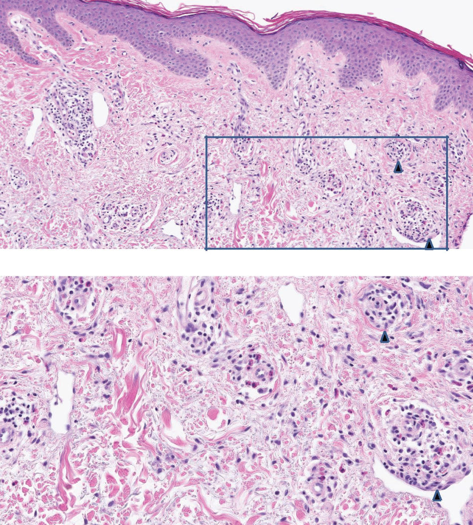
Punch biopsies of the skin were collected, and a sample from the left lower back confirmed the presence of ALHE (Figures 1 and Figure 2). A histologic examination of this sample revealed the presence of circumscribed vessels with enlarged endothelial cells in the dermis, surrounded by inflammatory cells, such as eosinophils, monocytes, and rare mast cells.
.
Figure 1 and Figure 2: Histologic examination from a punch biopsy of skin under hematoxylin and eosin (H&E) stain at 400x (Figure 1) and 800x (inset, Figure 2) magnification revealed a circumscribed dermal collection of vessels with plump endothelial cells. The vessels were surrounded by an inflammatory infiltrate composed of mononuclear cells, eosinophils, and rare mast cells. Two of these are marked with black arrowheads in both figures. Courtesy of Dr. David Jones, Albany Medical Center, Albany, NY, and Dr. Hal Schaffer, Hudson, NY.
View Figure 1 and Figure 2
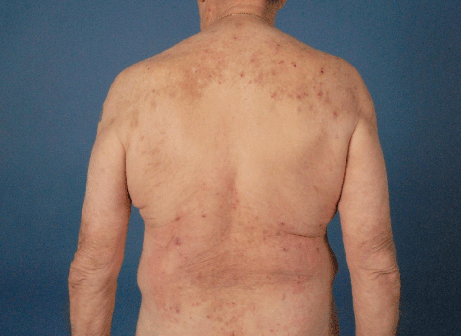
Four months later, once prednisone had again been tapered off and the rash had largely resolved, adalimumab was resumed at its previous dose. Over the course of ten months, the patient's rash recurred, involving erythematous papules at his back, buttocks, and left leg (Figure 3). Adalimumab was again discontinued in favor of etanercept 50 mg subcutaneously once weekly. Since the patient's joint pains were not well-controlled on etanercept, it was switched to certolizumab 400 mg subcutaneously every other week three times, then 200 mg subcutaneously every other week as the maintenance dosage and frequency. The patient has not had recurrence of a papular rash while on etanercept and certolizumab.
.
Figure 3: Erythematous rash, some of which featured papular lesions, involving the upper and lower back.
View Figure 3
Discussion
As the use of anti-TNF-α inhibitors has grown, an increasing number of drug-induced autoimmune diseases have also been reported. This case represents the first reported case of ALHE secondary to adalimumab use. Vasculitides have been the most commonly reported autoimmune complications, with various different cutaneous small-vessel vasculitides noted [2]. One review has noted the association of Sweet's syndrome and neutrophilic eccrine hidradenitis with adalimumab, the medication used for the patient in this case. Furthermore, palmoplantar pustular psoriasis has been associated with etanercept use, while leukocytoclastic vasculitis, pustular folliculitis, and psoriasiform dermatitis have been associated with infliximab use [3].
These findings demonstrate that while anti-TNF-α inhibitors have therapeutic effects in some patients, they in fact induce pathological processes in others [4]. While it is unclear exactly why this occurs, one hypothesis holds that immune complexes form between TNF-α and anti-TNF-α inhibitors, generating a cytotoxic immune reaction in the skin that results in various erythematous or lichenoid lesions [4]. Another hypothesis holds that inhibition of TNF-α alters the balance in the production of TH1 and TH2 cytokines, promoting the expression of type 1 interferons that then lead to the vasculitis reactions [2].
Of note, a United States Food and Drug Administration (FDA) study revealed that leukocytic vasculitis resolved when patients stopped using their anti-TNF-α inhibitors, with two-thirds of them suffering relapses with the resumption of the same inhibitors [5]. While the patient in this case had biopsy-confirmed ALHE instead of a vasculitis, he also demonstrated a relapse when adalimumab was resumed, suggesting that the hypotheses linking anti-TNF-α inhibitors to vasculitides may also be extended to explain the pathogenesis of ALHE.
Case reports of ALHE thus far have demonstrated idiopathic origin, and not an association with medications such as anti-TNF-α inhibitors. For example, one case report noted a 30-year-old female patient who presented with peripheral joint polyarthralgias and soft, large nodules on the extremities [6]. Histological examination in that case demonstrated eosinophilia and lymphocytosis without parasitic organisms, fibrinoid necrosis, or vasculitis. Altogether, the findings suggested the presence of ALHE [6]. The polyarthralgias and nodules subsided with administration of prednisone and diethylcarbamazine [6]. In our case, in accordance with The Naranjo criteria, ALHE was determined to be definitely secondary to administration of adalimumab [7].
Interestingly, the patient's rash did not recur with the use of other anti-TNF-α inhibitors, namely etanercept and certolizumab. The different structures of adalimumab, etanercept, and certolizumab may help explain why this is the case. Adalimumab, the first fully human-derived monoclonal antibody approved by the FDA, consists of the constant fragment (Fc) and antibody-binding fragment (Fab) of a typical human monoclonal antibody. However, the Fab component has murine-derived TNF-binding epitopes to help it bind to TNF-α. Etanercept, on the other hand, is a fusion protein that is composed of two extracellular domains of human soluble TNF receptor p75, bound directly to the Fc component of a human monoclonal antibody for stabilization, but without the Fab component. Certolizumab is unlike other anti-TNF-α inhibitors in that instead of being composed of the Fc of a humanized monoclonal antibody, it is composed of the antibody-binding fragment (Fab), which is conjugated to polyethylene glycol (PEGylated) to stabilize it. Since the formation of immune complexes depend on the presence of the Fab, and in many cases the presence of Fc, as well, the fact that etanercept (lacking Fab) and certolizumab (lacking Fc) did not lead to ALHE or another autoimmune reaction in this patient weakens support for immune complex formation as the etiology for the development of ALHE.
Conclusion
While ALHE has previously been reported to be idiopathic in origin, this case demonstrates the presence of ALHE in association with use of the anti-TNF-α inhibitor, adalimumab, as well as resolution with removal of this anti-TNF-α inhibitor. Prescribers of such medications should remain vigilant for the development of papular rashes and nodular lesions in patients who use them, and may consider skin biopsies to help determine whether ALHE may be the underlying process for these. The development of ALHE in response to anti-TNF-α inhibitor use is not only important for pharmacovigilance, but also useful in further understanding the pathogenesis of ALHE.
References
-
Mankekar G, Chainani GN, Bhatt C, Sha TM (2000) Angiolymphoid hyperplasia with eosbvophilia-case report. Indian J Otolaryngol Head Neck Surg 52: 167-168.
-
Sokumbi O, Wetter DA, Makol A, Warrington KJ (2012) Vasculitis associated with tumor necrosis factor-α inhibitors. Mayo Clin Proc 87: 739-745.
-
Hawryluk EB, Linskey KR, Duncan LM, Nazarian RM (2012) Broad range of adverse cutaneous eruptions in patients on TNF-alpha antagonists. J Cutan Pathol 39: 481-492.
-
Moustou AE, Matekovits A, Dessinioti C, Antoniou C, Sfikakis PP, et al. (2009) Cutaneous side effects of anti-tumor necrosis factor biologic therapy: a clinical review. J Am Acad Dermatol 61: 486-504.
-
Mohan N, Edwards ET, Cupps TR, Slifman N, Lee JH, et al. (2004) Leukocytoclastic vasculitis associated with tumor necrosis factor-alpha blocking agents. J Rheumatol 31: 1955-1958.
-
Kaushik P, Malaviya AN, Makar R (2000) Angiolymphoid hyperplasia with eosinophilia (ALHE): an uncommon condition with arthritis, subcutaneous nodules and eosinophilia. Clin Exp Rheumatol 18: 648-649.
-
Naranjo CA, Busto U, Sellers EM, Sandor P, Ruiz I, et al. (1981) A method for estimating the probability of adverse drug reactions. Clin Pharmacol Ther 30: 239-245.
