
International Journal of Oral and Dental Health
Third Molar Transplantation: A Case Report
BENTO Gabriela, HADDAD Marcela Filié* and Mariano Ronaldo Célio
Federal University of Alfenas, Brazil
*Corresponding author: Marcela Filié Haddad, School of Dentistry, Federal University of Alfenas, Gabriel Monteiro da Silva Street, 714, Center, Alfenas - MG - CEP 37.130-000, Brazil, Tel: (35) 3299-1460, E-mail: marcela.haddad@unifal-mg.edu.br
Int J Oral Dent Health, IJODH-1-022, (Volume 1, Issue 4), Case Report; ISSN: 2469-5734
Received: October 26, 2015 | Accepted: November 23, 2015 | Published: November 18, 2015
Citation: Filié BGHM, Célio MR (2015) Third Molar Transplantation: A Case Report. Int J Oral Dent Health 1:022. 10.23937/2469-5734/1510022
Copyright: © 2015 Filié BGHM, et al. This is an open-access article distributed under the terms of the Creative Commons Attribution License, which permits unrestricted use, distribution, and reproduction in any medium, provided the original author and source are credited.
Abstract
Nowadays, implant-supported prostheses has been evidenced and shown to have excellent results. One of the reasons for dental treatments is to maintain natural healthy functional teeth throughout the lifespan. However, when this is not possible, dental autotransplantation can be an option. This method has some advantages when compared with other ways of oral rehabilitation such as avoiding jaw development changes, representing a conservative treatment. It could be called "biological prosthesis". Therefore, autotransplantation is a good option for oral rehabilitation and it can be done in one or two appointments. In addition, autotransplatation has a great cost benefit and have favourable success rates when compared to integrated bone implants. A 17 year old healthy female patient presented at the Surgery Clinic of Federal University of Alfenas with a Dentist referral to extract tooth 37- left lower second molar, due to its huge crown destruction. During the clinical exam, it was noticed that tooth 38 was not erupted. Thus, the extraction of tooth 37 was done and, in another appointment, tooth 38 was also extracted and transplanted to the receptor alveolus. Clinical and radiographic follow ups showed good results.
Keywords
Transplantation, Molar, Third, Surgery, Oral
Introduction
Dental autotransplantation or autogenous tooth transplantation (ATT) was first reported in 1951 [1]. Autotransplantation involves transferring a tooth from its alveolus to another site in the same person [2]. Autogenous tooth transplantation is indicated in cases of dental-alveolar trauma, extensive caries with root involvement, tooth agenesis, iatrogenic complications and when the patient is not able to afford a prosthetic or implant rehabilitation [3]. It is also known that transplantation has a key role in the replacement of young patients' missing teeth [4]. Osseointegrated implants are generally contraindicated for young patients who have developed alveolar bone once as it can cause infraocclusion [5].
The literature reports excellent success rates following tooth transplantation when the appropriate protocol is followed. Andreasen found 95% and 98% long-term survival rates for incomplete and complete root formation of 370 transplanted premolars observed over 13 years [6]. Lundberg and Isaksson had success in 94% and 84% of cases for open and closed apices respectively in 278 autotransplanted teeth over 5 years. Kugelberg achieved success rates of 96% and 82% for 45 immature and mature teeth transplanted into the upper incisor region over 4 years. Cohen showed success in the ranges of 98-99% over 5 years and 80-87% over 10 years with transplanted anterior teeth with closed apices. Nethander found 5-year success rates of over 90% for 68 mature teeth transplanted with a 2-stage technique [7]. Josefsson found 4-year success rates of 92% and 82% respectively for premolars with incomplete and complete root formation [8]. These consistently high success rates are a contrast to the variable results reported in many older studies. Schwartz and others yielded success rates of only 76.2% at 5 years and 59.6% at 10 years [9]. Similarly, Pogrel found that his success rate for 416 autotransplanted teeth was 72% [10]. However, other investigators of that era had more positive results. Kristerson for example, obtained a success rate of 93% when 100 auto-transplanted premolars were observed for a mean of 6.3 years [11].
Tooth autotransplantation is a viable option for replacing a missing element because it can function as a normal tooth when the transplantation is successfully held [6]. A transplanted third molar is able to maintain natural space, alveolar bone volume, and the morphology of the alveolar ridge through proprioceptive stimulation [5]. Although the indications for autotransplantation are narrow, careful patient selection coupled with an appropriate technique can lead to exceptional esthetic and functional results. One advantage of this procedure is that placement of an implant-supported prosthesis or other form of prosthetic tooth replacement is not needed.
The success of this procedure depends on the periodontal ligament integrity, surgical expertise, trauma extension and the period of extra-alveolar tooth exposure [7]. Additional factors that influence the success of tooth transplantation include periodontal lesions and acute infection absence in the recipient socket [8]. The purpose of this article was to report a case of autotransplantation of the left lower third molar to replace the left lower second molar performed through two-stage surgical technique.
Case Report
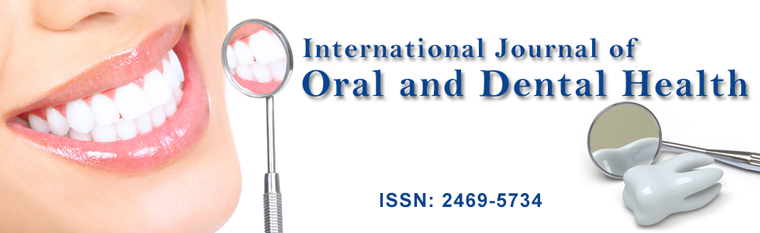
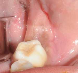
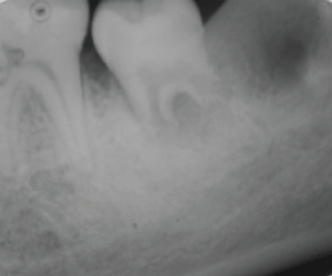
A healthy 17 year old female patient showed up at the Surgery Clinic of Federal University of Alfenas (UNIFAL-MG) Surgery Clinic. She had a Dentist referral to extract the tooth 37- left lower second molar, which had no possibility of restoration due to its huge crown destruction (Figure 1).
It was observed during the Clinical exam that the left lower third molar [3,8] was in good condition and an autotransplantation could be done. At that time, we suggested the autotransplantion to the patient so that she would not be toothless.
Before conducting the Surgery, it was recommended to the patient to take some medicines: analgesic (Lisador™ 500 mg); anti-inflamatory (Dexamethasone 4 mg) and antibiotic (Amoxicillin 500 mg). These medicines were taken 1 hour before the surgery. Also, a chlorexidine digluconate mouthwash (0.12%) was prescribed to use for 1 minute, 3 times a day, for 10 days, starting 3 days before the surgery.
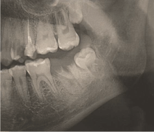
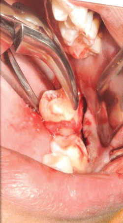
The procedure was done in two stages: 1- Extraction of the tooth 37 and alveolar preparation (Figure 2 and Figure 3).
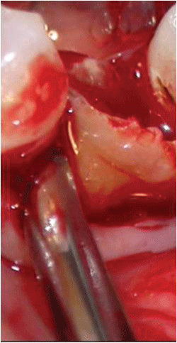
2- After 14 days, the edges of the wound were revived and the extraction of the tooth 38 (which would be transplanted), was held. However, the pericoronal hood was maintained together with the tooth (Figure 4 and Figure 5).
.
Figures 5: Atraumatic extraction of the tooth 38, the pericoronal hood was maintained together with the tooth.
View Figures 5
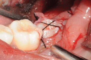
3- The tooth was inserted into the receiver socket and a "X" suture, using silk yarn, was made to keep it firm (Figure 6).
.
Figures 6: The tooth was inserted into the receiver socket and a "X" suture was made.
View Figures 6
Some medicines were prescribed after the surgery: analgesic (Lisador™ 500 mg) only in case of pain, ant-inflammatory, (Dexamethasone 4 mg) 24 hours after the Surgery and antibiotic (Amoxicillin 500 mg) for 10 days.
Also, it was recommended to the patient to eat only cold food in the first two days, then from the third day soft foods could be eaten. Furthermore, the patient was informed to return after 7 days and not to chew on the side of the wound.
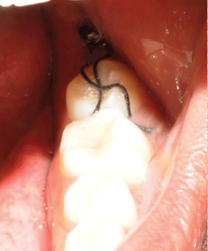
Seven days later the patient returned to remove the suture. At that time, the wound was healing well, the gums around the wound were healthy and there was no complaint from the patient (Figure 7 and Figure 8).
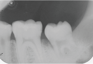
Fourteen days later, the transplanted tooth was in good condition. In the 34th day, continued root formation was observed (Figure 9 and Figure 10). After 4 months and 24 days, the transplanted tooth continued to be healthy as well as the gums around it. Also, the patient showed good oral hygiene. By using a radiographic exam, the continued root formation was seen (Figure 11).
.
Figures 11: Radiograph of the autotransplanted tooth 4 months and 24 days later.
View Figures 11
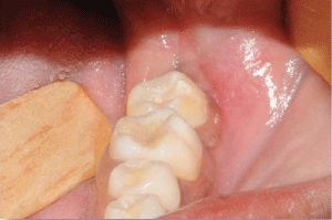
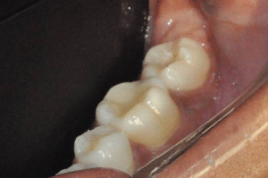
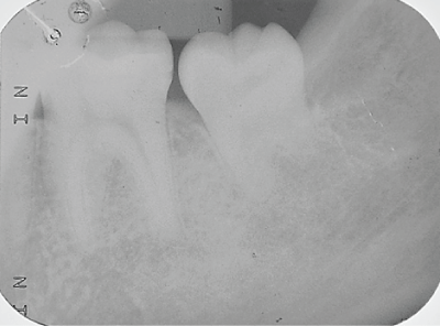
The transplanted tooth and the gums were healthy 1 year after the surgery. Moreover, the patient did not complain and said she was satisfied with the result (Figure 12 and Figure 13).
.
Figures 12: One year after the surgery. The transplanted tooth and the gum were healthy.
View Figures 12
.
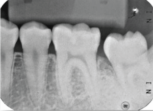
Figures 13: One year after the surgery. The radiograph shows the root formation.
View Figures 13
Discussion
The prognosis of an autotransplanted tooth is influenced by pre and post-operative conditions that are recognized as prognostic factors. Clinical studies have reported on teeth transplantation with incomplete roots and focused on factors such as development and eruption stage of the donor tooth as well as in root development, pulpal healing and root resorption of the transplanted tooth [11-14] The tooth of the patient had almost 2/3 of formed root, which was a good factor for the success of the treatment.
The factors that lead to success have been extensively investigated. The most significant determinant for survival of the transplant is the continued vitality of the periodontal membrane. In cases where the periodontal ligament is traumatized during transplantation, external root resorption and ankylosis is often noted [1,13]. In this case reported the extraction of the tooth was done in an atraumatic way, and the pericoronal follicle was preserved in order to preserve the periodontal ligament and the root cementum. Atraumatic extraction preserves the root structure, as well as influencing the outcome [10].
The experience of the surgeon also affects the success because this procedure is technique-sensitive. Although retention of the tooth and restoration of the edentulous space is the desired outcome for patients, more specific parameters have been used to measure the health of the surviving transplant. These parameters include marginal periodontal attachment, mobility, pain, root resorption, root development, sensitivity to percussion, gingival pocket depth, presence of gingivitis, and presence of fistulae [6,7]. In this case, there were no complaints of postoperative pain, excessive swelling, and dental mobility. Late postoperative radiographs showed no root resorption. Vertical and horizontal percussion tests showed appropriate responses. The depth of the gingival sulcus showed measures within normal standards.
The two-stage surgical technique used in the present case allowed observing that inflammatory root resorption was not present and periodontal tissues normally show aspects. We believe that the surgical technique used allowed to improve nutrition and preserve cell activity in periodontal tissues. Nethander et al. [7], Katayama et al. [15] and Ferreira et al. [16] suggested that teeth that be transplanted to the sockets with regenerative tissues, may reduce the root resorption and the circulation and innervations recover the original pulp tissue, and dentin development continues after transplantation of immature teeth. The one-stage surgical technique can increase the extra-alveolar time of the tooth that will be transplanted. During a prolonged extra-alveolar period, the pulp suffers and necrotizes. This indicates that increased handling of the transplant in attempts to adapt the tooth to the socket represents a risk of contamination with bacteria and damages the architecture and function of the pulp and periodontal ligament. To prevent this, we made the transplantation to a recipient bed in which the tissue was under regeneration as described by Nethander et al. [7], where the recipient bed is prepared surgically prior to transplantation and allowed to heal for 5 days. This allows in-growth and early maturation of granulation tissues into the wound, into which the transplanted teeth come in direct contact, improving the revascularization.
So, this case report was considered successfully because there was no root reabsorption or painful symptoms. In addition, there was normal periapical healing and the root growth continued without any inflammatory pulpal changes. The masticatory function of the transplanted tooth was satisfactory, the lamina dura appeared normal on radiographs and the gingival color and contour was in very good condition.
Conclusion
Tooth transplantation is a good way to treat dental loss, showing high success rates, low morbidity and low cost when compared to dental implants and prosthesis. Furthermore, it allows the transplanted tooth to remain healthy as well as its supporting tissues.
References
-
Miller HM (1951) Transplantation of teeth. N Y State Dent J 17: 382-386.
-
Frenken JW, Baart JA, Jovanovic A (1998) Autotransplantation of premolars. A retrospective study. Int J Oral Maxillofac Surg 27: 181-185.
-
Clokie CM, Yau DM, Chano L (2001) Autogenous tooth transplantation: an alternative to dental implant placement? J Can Dent Assoc 67: 92-96.
-
Thomas S, Turner SR, Sandy JR (1998) Autotransplantation of teeth: is there a role? Br J Orthod 25: 275-282.
-
Mendes RA, Rocha G (2004) Mandibular third molar autotransplantation--literature review with clinical cases. J Can Dent Assoc 70: 761-766.
-
Andreasen JO, Paulsen HU, Yu Z, Bayer T (1990) A long-term study of 370 autotransplanted premolars. Part IV. Root development subsequent to transplantation. Eur J Orthod 12: 38-50.
-
Nethander G (1994) Periodontal conditions of teeth autogenously transplanted by a two-stage technique. J Periodontal Res 29: 250-258.
-
Josefsson E, Brattstrom V, Tegsjo U, Valerius-Olsson H (1999) Treatment of lower second premolar agenesis by autotransplantation: four-year evaluation of eighty patients. Acta Odontol Scand 57: 111-115.
-
Schwartz O, Bergmann P, Klausen B (1985) Autotransplantation of human teeth. A life-table analysis of prognostic factors. Int J Oral Surg 14: 245-258.
-
Pogrel MA (1987) Evaluation of over 400 autogenous tooth transplants. J Oral Maxillofac Surg 45: 205-211.
-
Kristerson L, Lagerstrom L (1991) Autotransplantation of teeth in cases with agenesis or traumatic loss of maxillary incisors. Eur J Orthod 13: 486-492.
-
Kahnberg KE (1987) Autotransplantation of teeth (I). Indications for transplantation with a follow-up of 51 cases. Int J Oral Maxillofac Surg 16: 577-585.
-
Tegsjo U, Valerius-Olsson H, Frykholm A, Olgart K (1987) Clinical evaluation of intra-alveolar transplantation of teeth with cervical root fractures. Swed Dent J 11: 235-250.
-
Northway WM, Konigsberg S (1980) Autogenic tooth transplantation. The "state of the art". Am J Orthod 77: 146-162.
-
Katayama A, Ota M, Sugito H, Shibukawa Y, Yamada S (2006) Effect of proliferating tissue on transplanted teeth in dogs. Oral Surg Oral Med Oral Pathol Oral Radiol Endod 101: e110-118.
-
Ferreira M, Botelho M, Lina C, Oliveiros B, Carrilho E (2010) Histological evaluation of periodontal regeneration in autogenous tooth transplantation in the dog: a comparison between one and two-stage surgical techniques, a pilot study. Dent Traumatol 26: 76-79.
