

International Journal of Pediatric Research
Treatment Approach in Teeth with Complicated Crown and Crown-Root Fracture: Three Case Reports
Mehmet Sinan Dogan1*, Izzet Yavuz1, Osman Atas1, Abdullah Emre Karaali1, Fatih Demirci2 and Abdulsamet Tanik3
1Department of Pediatric Dentistry, Faculty of Dentistry, Dicle University, Diyarbakir, Turkey
2Department of Prosthodontics, Faculty of Dentistry, Dicle University, Diyarbakir, Turkey
3Department of Periodontology, Faculty of Dentistry, Dicle University, Diyarbakir, Turkey
*Corresponding author: Mehmet Sinan Dogan, Department of Pediatric Dentistry, School of Dentistry, Dicle University, 21280 Diyarbakir, Turkey, E-mail: dtlider@hotmail.com
Int J Pediatr Res, IJPR-1-006, (Volume 1, Issue 2), Case Series; ISSN: 2469-5769
Received: September 02, 2015 | Accepted: October 06, 2015 | Published: October 09, 2015
Citation: Dogan MS, Yavuz I, Atas O, Karaali AE, Demirci F, et al. (2015) Treatment Approach in Teeth with Complicated Crown and Crown-Root Fracture: Three Case Reports. Int J Pediatr Res 1:006. 10.23937/2469-5769/1510006
Copyright: © 2015 Dogan MS, et al. This is an open-access article distributed under the terms of the Creative Commons Attribution License, which permits unrestricted use, distribution, and reproduction in any medium, provided the original author and source are credited.
Abstract
Tooth injuries constitute an integral part of clinical dentistry. Complicated crown fracture and crown-root fracture are the most common injury in the permanent dentition. The success of treatment and prognosis of the traumatized tooth depends on accurate diagnosis and treatment procedures and materials. The aim of the case series is to present rehabilitation of the teeth with crown-root fracture as restorative and prosthetic by glass-fiber posts. 9, 12 and 13 years old three patients were referred to Dicle University Faculty of Dentistry, Department of Pediatric Dentistry, results of the clinical and radiological evaluations showed that 9 years old patient with tooth 11, 12 years old patient with tooth 11 and 13 years old patient with tooth 21 needed root canal treatment and glass fiber post application before the coronal restoration because of excessive loss of hard tissue. As a result, glass fiber posts were applied to these cases after root canal treatment, and patients were followed for two years clinically.
Keywords
Tooth fractures, Cone beam computed tomography, Fiberglass reinforced polymers
Introduction
Dental injuries that occur following dental trauma are cases that need urgent approaches in dentistry. Dental injuries may exist as a result of falling, crashing, traffic accidents and sportive injuries [1]. The risk of traumatic dental injury is higher in children with hyperactive and presence of malocclusions further increases the likelihood of dental injuries [2].
It has been reported that, dental injuries occur between the 2-4 ages in deciduous dentition period and between 8-10 ages at permanent dentition period [3]. Tooth and oral tissue injuries following trauma may cause psychological, aesthetic and psychologic problems. Both children and families are affected negatively because of these problems [2,4].
Dental trauma can vary from a minor enamel fracture to extensive maxillofacial injury involving the supporting structures and displacement or avulsion of teeth [5]. Anterior teeth fractures are very common in children and adults following trauma. Providing the aesthetic rehabilitation at fractured anterior teeth is a complicated case for the dentists. Ensuring a successful treatment at fractured anterior teeth related to the size of fractured part, condition of pulp, remaining tooth part, availability of fractured part and occlusion [6].
The complicated crown-root fractures may occur between the rate of 2-13% following the trauma and the fractured part includes enamel, dentin and pulp. Complicated crown fractures are mostly occur in upper central incisors [7].
They usually occur as a result of horizontal forces. The fracture line starts from the crown and proceed to the subgingival area [8]. The treatment options of complicated crown fractures changes related to the position and level of the fracture line replacement amount of tooth structures, type of occlusion and prognosis of the tooth. According to these conditions are used composite restorations and post-core restorations. Restorations following the extraction of the teeth, implant or fixed prosthetic restorations; Restorations following gingivectomy or osteotomy, orthodontic or surgical extrusion may be performed [8,9]. If the remaining crown length is enough for the restoration, exposed pulp tissue must be protected with calcium hydroxide and mineral trioxide aggregate [7]. It is claimed that; fragility rate of teeth following the root-canal treatment increase. The reason of increased fragility rate of teeth with root-canal treatment is having less mineralized tissue, loss of water, internal and external tissue loss, chemical materials that used while root-canal treatment and affects of medicaments that performed at the inside of root- canal [10].
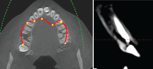
Root fractures are diagnosed with clinical and radiological examinations. The evaluation of mobility, percussion sensitivity, palpation sensitivity of soft tissue, crown length are performed while clinical examination. The fracture line must be followed carefully while radiographic evaluation. More than one periapical radiographs must be taken from the different angles in order to diagnose the fracture line. In addition, occlusal radiographic may help the diagnose [1]. But, if the x-rays do not correspond to fracture line or if the superposition of anatomic tissues or filling materials occurs, traditional radiographic may be inadequate. Recently, it has been reported that cone beam computed tomography (CBCT) is efficient for the diagnosis of root fractures [11].
Post-core restorations may performed for the rehabilitation of crown-root fractures. The purpose of post-core applications is to increase resistance of teeth with root-canal treatment against functional forces [12].
The aim of the case series is to present rehabilitation of the teeth with crown-root fracture as restorative and prosthetic by glass-fiber posts.
Material and Method
Case 1
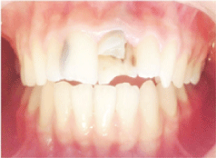
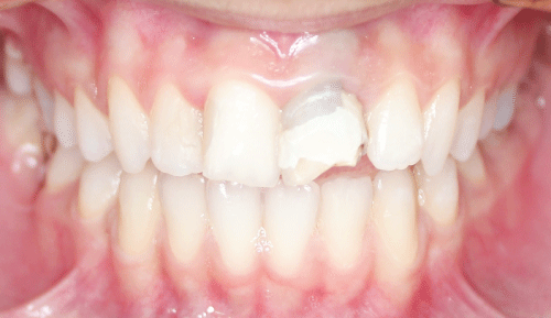
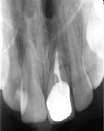
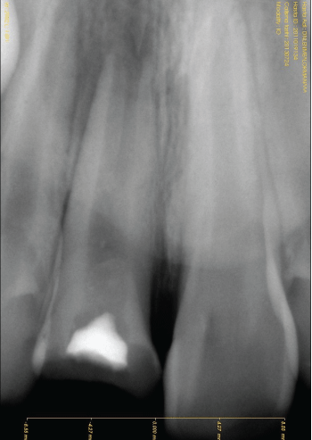
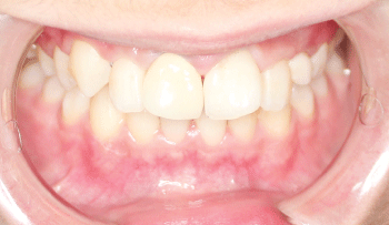
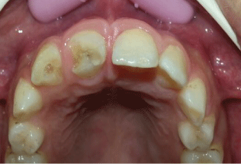
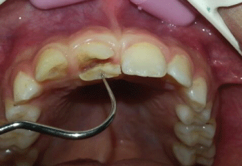
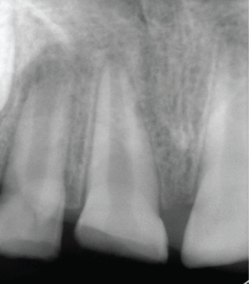
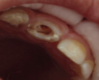
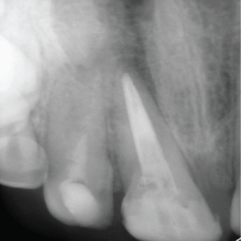
A 13-year-old systemically healthy child applied to Dicle University Faculty of Dentistry Department of Pediatric Dentistry with fracture at 21 numbered tooth as a result of hitting to a hard object while playing at park. It has been learned from the anamnesis that, 4 years ago root-canal treatment was performed to the same tooth following a trauma. Following the clinical and radiological examinations, a fracture was diagnosed which starts from the crown and proceed to the root (Figure 1-4).
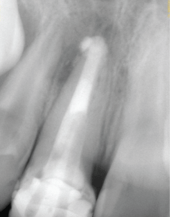
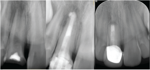
Cone beam computed tomography was taken from the patient for the certain diagnosis after receiving approval from the parent. As a result of the evaluation of the tomographic views, an oblique fracture was diagnosed which starts from the crown and proceed until the midline of the root (Figure 3). The patient and the family were informed about the treatment options and the approval received from the family. First of all, before the post-core restoration 1mmgingiva tissue was removed from the vestibule part of 21 numbered tooth by 15 numbered scalpel (Shandong Sinorgmed Co., Ltd. Shandong, P.R.C).
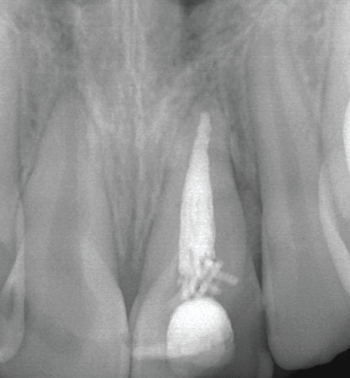
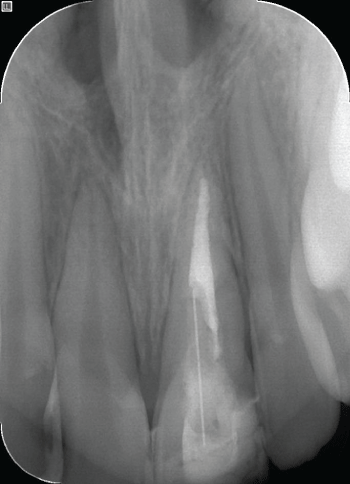
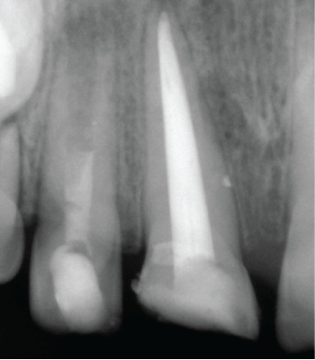
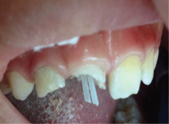
Thus, the gingival level of two upper central incisors regulated symmetric. Following session, the 2/3 part of gutta-percha was removed from the root canal by a drill (snowpost-16,carbotec, Ganges, France) in order to place the post material. The inside of the canal and the enamel-dentin tissues of the tooth etched by 37% phosphoric acid (3M Scotchobond; 3M ESPE, St. Paul, Minn, ABD) 15 seconds. Following the etching procedure, tooth was rinsed with water in order to remove the acid. The primer and bond of self-etching adhesive (Clearfil SE Bond, Primer; Kuraray Co. Ltd, Osaka, Japan) was applied to the tooth. Than the A and B parts of dual-cure adhesive material (Panavia F, Kuraray Co Ltd, Osaka, Japan) were taken equally and mixed 20 seconds. Than it was applied into the canal with a lentulo (Mani Inc, Tochigi-Ken, Japan) The glass fiber post (snowpost-16, carbotec, Ganges, France) was covered with the adhesive material and was placed with finger pressure. The light (Curing Light XL 3000; 3M, St Paul, MN, ABD) was performed 40 seconds for the polymerization of dual cure adhesive. The polymerization was completed with performing Oxyguard II gel (Kuraray, Osaka, Japan) 3 minutes. The long coronal part of fiber post was removed and the restoration was completed with hybrid composite resin (Clearfil AP-X, Kuraray, Osaka, Japan) (Figure 5). After the post application, periapical radiography was taken from the 21 numbered tooth (Figure 6).
.
Figure 6: The periapical radiography of 21 numbered tooth following fiber post application.
View Figure 6
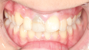
The fracture of the tooth was taken into consideration and thus, metal-ceramic restoration was planned. The preparation of anterior teeth was performed proper to metal ceramic preparation techniques and champher marginal end design was prepared. While making impression, retraction cord (Stay-put, Medium; Roeko, Langenau, Germany) was used in order to decrease gingival fluid and then a type silicon impression material was used for impression (A-Silicone Elite HD+, Zhermack, Rovigo, Italy). In order to enhance the aesthetic view, laser sintering method was performed to the metal substructure of prosthesis. Before the colour decision, polishing was performed to the anterior teeth of patient. The colour decision was performed with the traditional technique by vita 3D Master (Vita Zahnfabrik, Bad Sackingen, Germany) colour scale. After the ceramic restoration prepared, it was cemented, restorations were cemented with zinc-polycarboxylate cement (Adhesor Carbofine Zinc polycarboxylate cement, Spofa Dental, Jicin, Czech Republic) (Figure 7 and Figure 8).
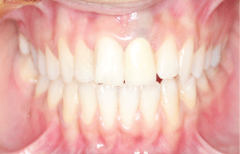
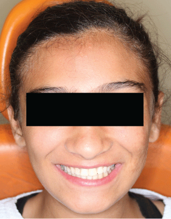
The patient was advised to come every 6 months for the control sessions. After the 2 years control period there were not any clinical and radiological problems (Figure 9).
Case 2
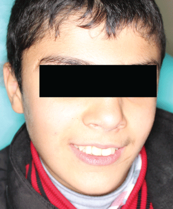
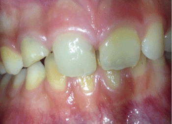
A 12-year-old systemically healthy male patient applied to Dicle University Faculty of Dentistry Department of Pediatric Dentistry with fracture at 11 numbered tooth as a result of hitting to a hard object while playing at park (Figure 10).
Following the root-canal treatment the same treatment procedure in case 1 was followed and the restoration was completed (Figure 11-14). We placed mineral trioxide aggregate (MTA) in large-open apices before the root canal treatment.
.
Figure 11: The radiographic view of 11 numbered tooth following fiber post placement.
View Figure 11
.
Figure 12: The intraoral view of 11 numbered tooth following fiber post application.
View Figure 12
But, the patient was pained after root-canal treatment. For this reason, we were performed apical resection. The patient was advised to come every 6 months for the control sessions. The radiographic examinations after 6 and 24 months did not show any pathological symptoms (Figure 15). The clinical examination also did not show any pathological symptoms.
Case 3
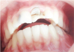
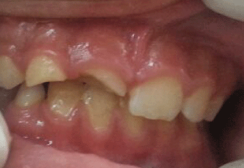
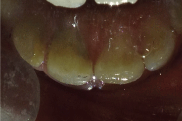
A 9-year-old male patient who was exposed to dental trauma has been applied to our clinic with the reason of fracture at upper right incisor. In anamnesis, any systemically disease was not determined and while questioning the trauma history, it was learned that the patient felt from the swing a month ago. In intraoral examination, complicated crown fracture which extends below the gingiva was observed (Figure 16-18).
In radiographic examination, it's observed that the apex of the tooth is open (Figure 19). In clinical examination, any sensitivity at percussion or palpation was not observed and the reaction to electrical vitality test was negative. Apexification with Frank method was performed at the same day because of open apex. After the endodontic access cavity, canal preparation was performed with H-files. After each preparation, The root canal was irrigated by 5,25% sodium hypochlorite between each file change and final irrigation was performed by saline solution.
Sterile paper points were used to dry root canal. Calcium hydroxide pasted (Sultan, USA) was applied to the root canal and glass ionomer cement (Fuji 9, Japan) was used as temporary filling material. The patient recalled after 3 weeks. Same procedures were repeated at second session. In radiographic examination which practiced after 2 months, apex was closed. At the same session, root canal was filled with AH Plus (Dentsply, Germany) and gutta-percha (Diadent, Korea) by lateral compaction technique (Figure 20). The patient was recalled after a week.
After that, the coronal 2/3 part of root canal filling material was emptied in order to place post by drills of the post system (Figure 21). Inside of the canal and enamel-dentin parts of the tooth were etched with 37% orthophosphoric acid (3M ESPE) and cleaned. Fiber post was placed into the root canal with adhesive resin cement and was polymerization 30 seconds by light device (Figure 22).
Then the long part of fiber post was cut and restoration was performed with resin composite (Filtex Supreme 3M ESPE USA). Polishing procedure was performed with polishing discs (Figure 23-25).
Discussion
Dental injuries usually causes severe problems that effect patients from the point of pain, function, aesthetic and psychological [13]. The basic factors that determine the injury level following dentoalveolar trauma are; the severity of trauma, type, direction, elasticity of the object, the absorption level of lips and other soft tissues, the quantity and quality of the tooth and jaw structures [14].
It has been reported that CBCT is an important option for the diagnosis of root fractures. The artefact occurrence decrease with CBCT. Some materials as gutta-percha, canal filling paste, and posts may cause artefacts. But, while diagnosing a root fracture, clinical and radiologic findings and symptoms must be evaluated together [15]. In case 1 because of the gutta-percha superposition, the fracture line could not determined clearly. That is why it was evaluated with taking 3D views by CBCT.
Post-core applications are usually performed in teeth with severe crown destruction. The purposes of restoration are; support, replacement and retention. Retention problem may occur at teeth with severe crown destruction and root-canal treatment. In recent years, in order to fix this problem, the usage of fiber supported post materials are commonly in use. The less invasive treatments are important for the young patients. The use of fiber posts is a conservative treatment plan for the tooth which is traumatized and with severe tissue destruction [16-18].
It has been stated that, homogen integrity occurs while using composite resin cement and core material with fiber posts [17]. When these materials are used together, they resist to chewing forces as monoblock and thus, the fracture resistance is reduced to minimal level [19-21]. Fiber posts have fiber bundles that have been buried in to a special resin matrix. In these bundles, fibers are in knitted form and powered with epoxy resin [22,23]. The physical properties of glass fiber posts are similar with dentin and composite resin. It is biocompatible; no risk for corrosion, the risk of fracture is really low. Post materials may be shortened according to the condition of the tooth and may removed easily from the canal with drill. These are the advantages [21,24,25]. Because of these advantages, in our cases, glass fiber post materials were used in order to strengthen the remaining toot structure, restore the severe structure loss of the crown and enhance the aesthetic view.
Calcium hydroxide has been broadly used to apexification at the root apex. For this purpose, this material is required the placement of long-term in the root canal. Consequently, root apex is treated with this material by induce formation of an apical hard tissue barrier. The formation of apical barrier is necessary to allow the filling of the root canal without risk of overfilling [26]. We are used to this method (Frank's method) for our third case in order to formation of an apical hard tissue barrier.
MTA is a bioactive material, which induces hydroxyapatite crystal formation on its surface when in contact with tissue fluids. This phenomenon is responsible for its biocompatibility, hard-tissue induction potential and sealing ability, features that make MTA an appropriate material for use as an apical plug in teeth with necrotic pulps and open apices [27]. In our second case, we were using the MTA as an apical plug in tooth with necrotic pulp and open root apices. However, the case was occurred pain after this process. Consequently, we have to performed apical resection.
Currently, MTA is believed by many authors as the material of choice for apical plug in cases of necrotic pulp and large-open apices. One of the major aims is its well sealing ability. But, the extrusion of this material to the periapical tissue can prevent with the repair process. Moreover, this substance has a high cost and to have difficulty applying the apical region, including the risk of extrusion to the periapical tissues, which could give rise to tissue injury and harm to the repair process [28]. If root -canal therapy fails, endodontic surgery may be required. Endodontic surgery involves a combination of curettage of infected tissue and removal of an infected or damaged root apex [29]. Because of these, we were performed apical resection for our second case.
It is possible to save the teeth with crown fracture or root fracture or both together for a long time following the root-canal treatment and post application and deciding to a proper restoration [30].
Glass fiber, polyethylene fiber and zircon post system are developed for restoration of fracture teeth which are occurred loss structure. The studies are reported that fiber post systems elasticity modulus is more closely to dentin compared with metal post system. For this reason, fiber post systems are more resistant to against stress [31]. Our third case, we are performed the endodontic treatment of tooth which occurred complicated crown- root fracture and immature by providing formation of apical barrier to allow the filling root canal by means of using to Frank method. We are used to glass fiber post system due to loss structure of tooth and the fractured part of tooth. Thus, we were providing to come to an adequate level functional and eliminate of aesthetic concern.
To protect the tooth tissue is always important if the tissue loss is severe. To obtain aesthetic, integrity of remaining tooth structure and for the rehabilitation of discoloration, prosthetic treatments may performed at post applications [32]. For this reason, in our cases, metal ceramic restorations were performed.
Conclusion
Trauma in permanent anterior teeth causes psychological stresses at children. Proper occlusion relation affects the success and prognosis of treatment at the cases with complicated root-crown fractures. After performing this type of treatment vertical root fractures may occur due to physical trauma, early occlusal contact, heavy and stressed chewing and iatrogenic reasons. This type of treatment may be contraindicated in deep bite and bruxism cases. Compatible relation between adjacent and opposed teeth obtains well prognosis. It's necessary to follow cases with complicated root-crown fracture in a regular period. If there is any complication, early diagnosis may be done and treatment may be performed.
References
-
Celikten B, Uzuntas CF, Safaralizadeh R, Demirel G, Sevimay S (2014) Multidisciplinary approach for the treatment of horizontal root-fractured maxillary anterior teeth. Case Rep Dent 2014: 472759.
-
Correa-Faria P, Paiva SM, Pordeus IA, Ramos-Jorge ML (2015) Influence of clinical and socioeconomic indicators on dental trauma in preschool children. Braz Oral Res 29: 1-7.
-
Ritwik P, Massey C, Hagan J (2015) Epidemiology and outcomes of dental trauma cases from an urban pediatric emergency department. Dent Traumatol 31: 97-102.
-
Sardana D, Goyal A, Gauba K (2014) Delayed replantation of avulsed tooth with 15-hours extra-oral time: 3-year follow-up. Singapore Dent J 35: 71-76.
-
Namdev R, Jindal A, Bhargava S, Bakshi L, Verma R, et al. (2014) Awareness of emergency management of dental trauma. Contemp Clin Dent 5: 507-513.
-
Murthy CS, Kumari A , Vora J, Kowsky D (2012) Bio-Aesthetic Management of Complicated Crown Fracture - A Case Report. International Journal of Contemporary Dentistry 2: 133-136.
-
Vadini M, De Angelis F, D'Amario M, D'Arcangelo C (2011) Direct pulp capping with an adhesive system in management of a complicated incisor fracture: a three-year follow-up case report. Giornale Italiano di Endodonzia 25: 162-167.
-
Akyuz SN, Erdemir A (2012) Restoration of tooth fractures using fiber post and fragment reattachment: Three case reports. Eur J Gen Dent 1: 94-98.
-
Fidel SR, Fidel-Junior RA, Sassone LM, Murad CF, Fidel RA (2011) Clinical management of a complicated crown-root fracture: a case report. Braz Dent J 22: 258-262.
-
Hubbezoglu I, Kustarci A, Bek B (2007) Management Of Crown And Root Fractures. In: Endodontically Treated Teeth with Laser Application: Case Reports. Cumhuriyet Universitesi Dis Hekimligi Fakultesi Dergisi 10: 44-49.
-
Jakobson SJ, Westphalen VP, Silva Neto UX, Fariniuk LF, Schroeder AG, et al. (2014) The influence of metallic posts in the detection of vertical root fractures using different imaging examinations. Dentomaxillofac Radiol 43: 20130287.
-
Kustarci A, Tugut F, Ozcoban H, Kirmali O, Zan R (2011) Effects Of Two Different Root Canal Obturation Techniques And Two Different Post Systems On Apical Leakage. Ataturk Uni Dis Hek Fak Derg 21: 94-101.
-
Tugut F, Unal M, Kapdan A, Demir H, Dogan OM (2009) Composite Restoration Supported With Glass Fiber Post. In: Case Complicated Crown Fracture: Report of a Case And 18 Month Follow Up. Dis Hek Fak Derg. 19: 207-212.
-
Ozel E, Altundal H (2006) Dentoalveolar And Perioral Soft Tissue Injuries. Ataturk Univ Dis Hek Fak Derg 1: 7-13.
-
Bernardes RA, de Moraes IG, Hungaro Duarte MA, Azevedo BC, de Azevedo JR, et al. (2009) Use of cone-beam volumetric tomography in the diagnosis of root fractures. Oral Surg Oral Med Oral Pathol Oral Radiol Endod 108: 270-277.
-
Yanikoglu N, Bayindir F (2003) Restorative Materyals and Properties Used in Build Up Post-core. Ataturk Univ Dis Hek Fak Derg 13: 39-47.
-
Ricketts DN, Tait CM, Higgins AJ (2005) Post and core systems, refinements to tooth preparation and cementation. Br Dent J 198: 533-541.
-
Peroz I, Blankenstein F, Lange KP, Naumann M (2005) Restoring endodontically treated teeth with posts and cores-a review. Quintessence Int 36: 737-746.
-
Dean JP, Jeansonne BG, Sarkar N (1998) In vitro evaluation of a carbon fiber post. J Endod 24: 807-810.
-
Boschian Pest L, Cavalli G, Bertani P, Gagliani M (2002) Adhesive post-endodontic restorations with fiber posts: push-out tests and SEM observations. Dent Mater 18: 596-602.
-
Kivanc BH (2006) Application of posts in endodontically treated teeth. AtatUrk Univ Dis Hek Fak Derg 1: 18-23.
-
Uzun G, Keyf F (2007) An Alternative to Conventional Post-Core Systems: Polyethylene Fiber Post. Hacettepe Dis Hek Fak Derg 2: 43-48.
-
cicek E, Parlak E, Tunga U (2012) Application of glass fibre post: two case reports and two years follow up. AtatUrk Univ Dis Hek Fak Derg. 22: 66-70.
-
cokUk N (2009) Application of esthetic posts. In: Endodontically treated teeth. AtatUrk Univ Dis Hek. Fak. Derg 19:124-130.
-
Mannocci F, Sherriff M, Watson TF (2001) Three-point bending test of fiber posts. J Endod 27: 758-761.
-
SOUZA Ronaldo Araujo, Silva-Sousa YT, Colombo S, Lago M, Duarte MA, et al. (2012) Healing of a tooth with an overinstrumented apex, extensive transportation and periapical lesion using a 5 mm calcium hydroxide apical plug: an 8-year follow-up report. Braz Dent J. 23: 608-611.
-
Garip H, Garip Y, Orucoglu H, Hatipoglu S. (2011) Effect of the angle of apical resection on apical leakage, measured with a computerized fluid filtration device. Oral Surg Oral Med Oral Pathol Oral Radiol Endod 111: 50-55.
-
Chala Sanaa, Abouqal Redouane, Rida Sana (2011) Apexification of immature teeth with calcium hydroxide or mineral trioxide aggregate: systematic review and meta-analysis. Oral Surgery, Oral Medicine, Oral Pathology, Oral Radiology, and Endodontology 112: 36-42.
-
Nosrat A, Nekoofar MH, Bolhari B, Dummer PM (2012) Unintentional extrusion of mineral trioxide aggregate: a report of three cases. Int Endod J 45: 1165-1176.
-
ozsezer E, Akman A (2009) The treatment of crown and intraalveoler Root Fracture of Right Maxillary First Molar (A Case Report). AU Dis Hek. Fak Derg 36: 109-114.
-
Qing H, Zhu Z, Chao Y, Zhang W (2007) In vitro evaluation of the fracture resistance of anterior endodontically treated teeth restored with glass fiber and zircon posts. J Prosthet Dent 97: 93-98.
-
ozdemir E, AgUloglu S (2006) A Crown restoration of fiber reinforced composite which is supported from the root canal. TUrkiye Klinikleri J Dental Sci 12: 123-126.
