

International Journal of Surgery Research and Practice
Laparoscopic Resection of a Large Mesenteric Cyst - A Case Report
Kim WFM Lambregts 1*, Willem M Deserno2 and Jeroen Heemskerk1
1Department of General Surgery, Laurentius Hospital Roermond, The Netherlands
2Departments of Radiology, Laurentius Hospital Roermond, The Netherlands
*Corresponding author: Kim WFM Lambregts, Department of General Surgery, Laurentius Hospital Roermond, Monseigneur Driessenstraat 6, 6043 CV Roermond, The Netherlands, Tel: 0475382222, E-mail: Kwfm.lambregts@student.maastrichtuniversity.nl
Int J Surg Res Pract, IJSRP-1-009, (Volume 1, Issue 1), Case Report; ISSN: 2378-3397
Received: August 31, 2014 | Accepted: October 01, 2014 | Published: October 03, 2014
Citation: Lambregts KWFM, Deserno WM, Heemskerk J (2014) Laparoscopic Resection of a Large Mesenteric Cyst - A Case Report. Int J Surg Res Pract 1:009. 10.23937/2378-3397/1410009
Copyright: © 2014 Lambregts KWFM, et al. This is an open-access article distributed under the terms of the Creative Commons Attribution License, which permits unrestricted use, distribution, and reproduction in any medium, provided the original author and source are credited.
Abstract
Mesenteric cysts are rare lesions occurring in the abdomen. These cystic lesions can be asymptomatic or present with a specific symptoms. The decision whether to perform open or laparoscopic surgery depends on different features of the cystic lesion. If the cyst is thought to be of benign origin, a laparoscopic resection can be performed. We present a case of a young female with a large mesenteric cyst of 28cm, resected with laparoscopic approach.
Keywords
Mesenteric cyst, Abdominal cyst, Case reports, Laparoscopic surger
Abbreviations
CT: Computer Tomography; MRI: Magnetic Tesonance Imaging; US: Ultrasonography
Introduction
Mesenteric cysts are rare cysts located in the mesentery of the small bowel or colon [1]. These cysts can remain asymptomatic, until they reach considerable size. We present a case of a 33 year old female, in whom a huge mesenteric cyst was coincidentally discovered during her first pregnancy. Eventually, she was operated using a laparoscopic approach.
Case Report
In 2006, a 26-year old female, pregnant with her first child, presented for a regular check-up. Gestational age of the child was nineteen weeks. During routine US, a space occupying lesion was discovered next to the uterus with a size of 15 x 12 x 6 cm, transonic, with bright echo density, initially diagnosed as a pathologic cystic lesion of the right ovary. However, as there were no other findings supporting a malignant diagnosis, tumor markers CEA, alfa-fetoprotein and CA125 were within normal range, and the intra-uterine pregnancy was intact, watchful waiting was indicated. Over twenty weeks later, the patient delivered a healthy female baby after a spontaneous, uncomplicated childbirth. Several weeks following delivery the patient was examined and was found to have normal internal genitalia; therefore, in February 2007 abdominal MRI with gadolinium was performed, showing a sharply demarcated additional structure, with a hypo-intense dimension on T1-weighed haste and highly hyper-intense dimension on T2 weighed haste, with no contrast uptake of gadolinium. It was defined a cystic lesion, most compatible with a benign mesenteric cyst (Figure 1). Therefore, the patient was referred to the surgeon in order to arrange total cyst resection by laparotomy. Due to personal circumstances, including a second pregnancy, the operation was postponed for many years. In 2013, the patient returned to department of surgery with complaints of symptomatic abdominal distention, specifically on the left side of the abdomen. These complaints have been present for a few months and are slowly progressive. She has no complaints of pollakiuria or urgency. By this time, she reached the age of 32 and had no accompanying symptoms such as weight loss or malaise. A CT scan was performed, showing progressive growth of the cystic mass, with compression of the bladder (Figure 2). Apart from the progressive growth, there were no signs of malignant transformation, invasion of other organs or lymfadenopathy.
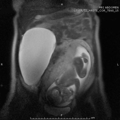
Figure 1: Coronal T2 Weighted Haste (TE/TR/FA=60ms./1100ms./150�)
MRI scan showing a large cyst of 21x11x16cm. The uterus is shifted to the
left by the cyst, while the small intestine is moved to the upper left quadrant.
At this time, the patient didn�t have any symptoms.
View Figure 1
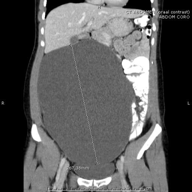
Figure 2: CT scan after 66 months demonstrates the progressively grown cyst, with a cranio caudal diameter of 27 cm with bladder compression. There was no relation between cyst, uterus and ovaries, during scan no lymphadenopathy could be demonstrated consistent with a large mesenteric cyst.
View Figure 2
A laparoscopic resection was performed, using a three-trocar-technique. The cyst with an estimated size of 30 x 15 x 20 mm was easily punctured and four liters of translucent fluid were aspirated. The cystic wall was resected from the mesenterium using Harmonic Ace (Johnson & Johnson, New Brunswick, USA), and was removed through a 12mm trocar in the left inguinal region (Figure 3). Inspection of the intra-abdominal region showed no residual cystic substance, bleeding or visible lesion to the intestines. Operating time was 72 minutes. The patient recovered well and was discharged from the hospital the next day. Clinical examination two weeks later revealed no abnormalities and the patient experienced no complaints. A post-resection CT-scan is not performed to confirm a complete resection, as there was no evidence of residual cystic substance in the abdomen during peroperative inspection. She was discharged from follow-up.
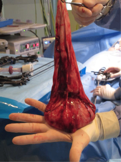
Figure 3: Peroperative image of resected cyst after aspiration of four liters of translucent fluid. The cyst was removed in toto through a 12mm trocar in the left inguinal region
View Figure 3
Microscopic examination revealed a cyst of enteric origin, with variable connective tissue and a lining that consists of single layer columnar- and cuboidal epithelium. No atypia or infiltrative features were discovered.
Discussion
Mesenteric cysts are rare cysts located in the mesentery of the small bowel, large intestine or, rarely, in the mesentery of the descending colon, sigmoid of rectum [1]. They are identified in about 1 in 100,000 hospital admissions among adults and up to 1 in 20,000 acute pediatric admissions [2,3]. These cysts can be asymptomatic, cause mild symptoms or result in an acute abdomen. Asymptomatic mesenteric cysts are usually discovered by coincidence using radiologic imaging such as Ultrasonography (US), Computed Tomography (CT) or Magnetic Resonance Imaging (MRI) for other reasons. If the cyst causes mild symptoms, patients usually seek medical attention. It is important to treat these cysts by resecting them, since there is a chance the cyst can lead to an acute abdomen, induced by torsion, rupture, infection or compressive effects on other organs, potentially resulting in obstruction [2].
Originally, mesenteric cysts were divided in four groups according to Bearhs (1950). This classification was based on etiologic and clinical features, and consisted of four groups: embryonic and developmental, neoplastic, traumatic and acquired and finally infective and degenerative [4]. Since 1950, more differences between mesotheliomas and lymphangiomas were appreciated, necessitating a new classification. In 2000, a new classification was proposed by de Perrot, based on the histopathologic features of the internal epithelium. This classification comprises six groups, namely lymphatic cysts, mesothelial cysts, cysts of enteric origin, cysts of urogenital origin, mature cystic teratoma (dermoid cyst) and non pancreatic pseudocysts [5]. This new classification is important, since every group has distinct clinical features. Lymphatic cysts can be divided in simple lymphatic cysts and lymphangiomas, whereas mesothelial cysts can be subdivided in simple mesothelial cysts, benign cystic mesothelioma and malignant cystic mesothelioma. Simple lymphatic and mesothelial cysts usually remain stable and asymptomatic over time, whereas lymphangiomas and benign cystic mesotheliomas can be aggressive and invasive [5]. Malignant cystic mesothelioma can present benign, which can be another reason to resect a mesenteric cyst once discovered.
Once a mesenteric cyst is established, radiologic imaging is used to determine the nature of the cyst, most often using US [3]. However, the definitive diagnosis can only be established after resection. To delineate the relationship of the cyst to adjacent organs, a CT scan has to be performed. After radiologic imaging, an operation can be planned. It is advisable to resect the mesenteric cyst, not only to avoid complications of rupture, torsion or obstruction, but also to prevent malignant transformation and growth in other organs [5]. The chance of a mesenteric cyst to be malignant is about 3% [5,6]. For many years, the operation of choice for resecting a mesenteric cyst was a laparotomy. However, in 1993, Mackenzie performed a laparoscopic excision [6]. This approach is increasingly used ever since.
The decision whether to perform open or laparoscopic excision of a mesenteric cyst depends on many features of this cyst. If an underlying malignancy is expected, laparotomy is the method of choice, since the tumor has to be removed in total, followed by debulking of the involved organs. Also, if the cyst is possibly haemorrhagic, inflammatory, infectious or parasitic, laparotomy might be preferred [6]. Furthermore, the experience of the surgeon with laparoscopy has to be taken into account. If the cyst is thought to be of benign origin, a laparoscopy can be performed. The peritoneal cavity should be assessed, along with complete resection of the cyst wall [7]. Although operating time is longer with laparoscopy compared with laparotomy, the advantages are high; minimal invasive techniques comprise less postoperative pain, shorter hospital stay, early return to daily life activities and better cosmetics [1]. Conversion to an open procedure may occur, due to major intra-abdominal adhesions, malignant or infectious appearance during laparoscopy, complications while resecting the cyst or because of size issues [6]. In this case, the cyst could be separated from the adjacent organs and be removed via a 12mm trocar in the left inguinal region.
References
-
Bhandarwar AH, Tayade MB, Borisa AD, Kasat GV (2013) Laparoscopic excision of mesenteric cyst of sigmoid mesocolon. See comment in PubMed Commons below J Minim Access Surg 9: 37-39.
-
Lee DL, Madhuvrata P, Reed MW, Balasubramanian SP (2013) Chylous mesenteric cyst: A diagnostic dilemma. See comment in PubMed Commons below Asian J Surg.
-
Mihmanli I, Erdogan N, Kurugoglu S, Aksoy SH, Korman U (2001) Radiological workup in mesenteric cysts: insight of a case report. See comment in PubMed Commons below Clin Imaging 25: 47-49.
-
Yoldemir T, Erenus M (2013) Fatty necrosis of a mesenteric cyst in a woman initially diagnosed with a large ovarian cystic mass. See comment in PubMed Commons below J Obstet Gynaecol 33: 534-535.
-
de Perrot M, Br�ndler M, T�tsch M, Mentha G, Morel P (2000) Mesenteric cysts. Toward less confusion? See comment in PubMed Commons below Dig Surg 17: 323-328.
-
Kurnicki J, Swiatkiewicz J, Wrzesinska N, Skorski M (2011) Laparoscopic treatment of a huge mesenteric pseudocyst - case report. See comment in PubMed Commons below Wideochir Inne Tech Malo Inwazyjne 6: 167-172.
-
Kwan E, Lau H, Yuen WK (2004) Laparoscopic resection of a mesenteric cyst. See comment in PubMed Commons below Gastrointest Endosc 59: 154-156.
