

Journal of Clinical Gastroenterology and Treatment
Unusual Mixed Presentations of Multiple Jejunal Diverticulae with Small Bowel Obstruction and Perforation - Case Report
Lillian Reza, Madeeha Aziz, Olga Tsiamita and Abraham Ayantunde*
Department of Surgery, Southend University Hospital, Prittlewell Chase, Westcliff-on-Sea, UK
*Corresponding author:
Abraham Ayantunde, Department of Surgery, Southend University Hospital, Prittlewell Chase, Westcliff-on-Sea, SS0 0RY, United Kingdom, Tel: 01702435555, Fax: 01702385856, E-mail: biodunayantunde@yahoo.co.uk
J Clin Gastroenterol Treat, JCGT-2-025, (Volume 2, Issue 2), Case Report; ISSN: 2469-584X
Received: February 05, 2016 | Accepted: May 24, 2016 | Published: May 26, 2016
Citation: Reza L, Aziz M, Tsiamita O, Ayantunde A (2016) Unusual Mixed Presentations of Multiple Jejunal Diverticulae with Small Bowel Obstruction and Perforation - Case Report. J Clin Gastroenterol Treat 2:025. 10.23937/2469-584X/1510025
Copyright: © 2016 Reza L, et al. This is an open-access article distributed under the terms of the Creative Commons Attribution License, which permits unrestricted use, distribution, and reproduction in any medium, provided the original author and source are credited.
Abstract
Jejunal diverticulae are a rare phenomenon that is usually asymptomatic but can present with chronic abdominal pain or as an acute abdomen when complicated by intestinal diverticulitis, obstruction, haemorrhage or perforation. The clinical presentation is often atypical and therefore diagnosis occurs at surgical intervention or occasionally during an imaging investigation. Surgical resection of the affected bowel segment with primary anastomosis is the preferred treatment of choice in complicated jejunal diverticulosis.
We present a case of 94-year-old lady with an atypical presentation of complicated jejunal diverticulosis. She presented with acute abdomen, signs and symptoms suggestive of acute small bowel obstruction in the absence of specific diagnostic features of complicated jejunal diverticulae on the plain abdominal X-ray and CT imaging. Exploratory laparotomy revealed multiple jejunal diverticulae with several concealed perforations at the mesenteric border, distal to which an impacted jejunal enterolith was thought to have caused the small bowel obstruction. She made an uncomplicated recovery following small bowel resection of the affected perforated segment and a primary anastomosis. This case highlights the unusual mixed presentations of this rare entity and the importance of considering small bowel diverticulosis as a possible differential of acute abdomen especially in the elderly.
Keywords
Jejunal diverticulosis, Jejunal perforation, Peritonitis, Small bowel obstruction, Imaging
Introduction
The incidence of jejunal diverticula at autopsy is between 0.3-1.3% [1]. They are usually located on the mesenteric border of the small bowel, and are believed to cause clinical problem in an acquired condition. The prevalence increases with age and most commonly presents in the sixth and seventh decades of life [2]. It is a rare condition and usually asymptomatic but can cause considerable morbidity and mortality in the elderly if complicated by haemorrhage, perforation, mechanical obstruction or diverticulitis [3]. Mechanical intestinal obstruction secondary to enterolith, adhesions, intussusception or volvulus or internal herniation is reported in 2.3-4.6% of cases [2]. Diagnosis of complicated jejunal diverticulosis is difficult due to its atypical and non-specific clinical presentation hence the diagnosis is often made at laparotomy.
We describe the case of a 94-year-old lady who presented with atypical and mixed presentations of complicated multiple jejunal diverticulosis and to highlight the need for a high index of suspicion.
Case Report
A 94-year-old Caucasian woman was initially admitted under the care of the medical team with a seven day history of recurrent vomiting followed by frequent loose motion. She had associated progressive abdominal pain and distension. Her past medical history included transient ischaemic attack (TIA), gout, bilateral hip replacements, hysterectomy and radiotherapy for endometrial carcinoma ten years previously. Prior to admission she was living alone and independent of all activities of daily living.
Physical examination revealed severe dehydration, a distended abdomen with generalised tenderness, and a soft reducible wide neck umbilical hernia. Active bowel sounds were noted. Digital rectal examination demonstrated an empty rectum.
Blood investigations showed a haemoglobin level of 13.3 g/dl, a white cell count of 22.5 × 10⁹/L, and C- reactive protein at 268 mg/L. Renal function on admission showed a urea level at 37.8 mmol/L and creatinine of 291 umol/L. Liver function tests were unremarkable apart from a low albumin at 29 g/L. An arterial blood gas analysis revealed metabolic acidosis with a pH of 7.31, lactate 3.98 and bicarbonate 20.9. Plain abdominal X-ray demonstrated significantly dilated loops of small bowel in keeping with small bowel obstruction.
She stayed under the acute medical team for 3 days with a presumed diagnosis of severe gastroenteritis with secondary acute kidney injury until we were involved in her care. Despite intravenous fluid replacement therapy and broad spectrum antibiotics her inflammatory markers remained high. She subsequently developed two episodes of feculent vomiting and absolute constipation 24 hours prior to surgical involvement.
.
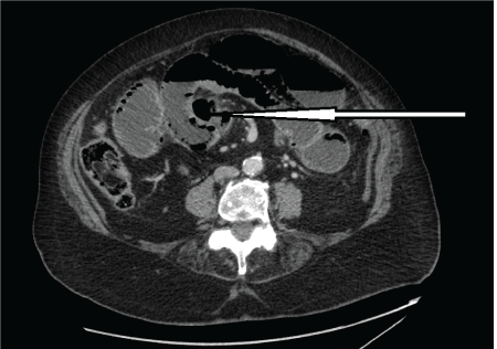
Figure 1: CT scan showing multiple jejunal diverticula and apparent air within the bowel wall. Large diverticula indicated by an arrow.
View Figure 1
.
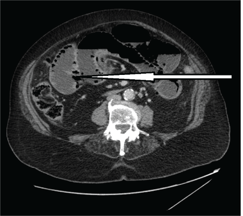
Figure 2: CT scan showing multiple jejunal diverticula mimicking pneumatosis intestinale. Multiple jejunal diverticula indicated by an arrow.
View Figure 2
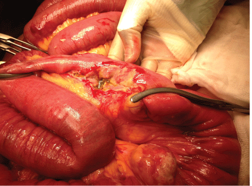
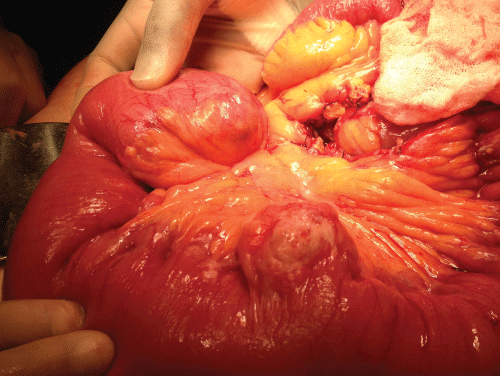
The differential diagnoses of acute small bowel obstruction and acute ischaemic bowel were entertained. An urgent CT scan of abdomen and pelvis showed a long segment of obstructed and possibly ischaemic dilated small bowel with pneumatosis intestinale (air within the bowel wall) (Figure 1 and Figure 2). There was an abrupt zone of transition at the junction of the proximal two thirds and distal one third of the small bowel. No free peritoneal air or fluid visualised. The patient and family were fully counselled on the risks and benefits of an emergency laparotomy, and given her fair premorbid state, decision was finally taken for emergency laparotomy. The findings at operation included a large obstructing enterolith within the proximal ileum, multiple jejunal diverticulae with three concealed perforated diverticulae walled off by bowel loops and omentum (Figure 3 and Figure 4). The whole of jejunum and proximal ileum were dilated and distal ileum was collapsed with cut off at the level of the impacted enterolith. There was also a near perforation at the level of enterolith impaction. The impacted enterolith was milked after a 40 cm segment of distal jejunum and proximal ileum involved with perforations were resected and a side-to-side TLC 75 stapled anastamosis was carried out. The histology of the resected specimen showed a 40 cm with perforations and evidence of peritonitis. There were focal mucosal ulcerations with abscess formation in small bowel at the sites of diverticular perforations.
.
Figure 3: Intraoperative findings of perforated jejunal diverticula and a large unperforated diverticulum.
View Figure 3
Patient made an uneventful postoperative recovery requiring an initial management in the surgical high dependency and total parenteral nutrition for the first five days post surgery. She was discharged home for subsequent follow-up in the surgical outpatient department.
Discussion
Small intestinal diverticulosis is a rare condition with a limited number of documented cases of complicated presentations. It was first described by Sommering in 1794 and later in 1807 by Sir Astley Cooper [3]. The prevalence of small intestinal diverticula on autopsy is between 0.06% to 1.3% [1], 80% of which are found in the jejunum, 15% in ileum and 5% in both [3].
There are a number of aetiopathogenetic explanations for jejunal diverticulosis and these are based on abnormalities of the smooth muscle and myenteric plexus. Jejunal diverticula are essentially pseudodiverticula, formed from mucosal and submucosal herniation through the muscular layer of the bowel wall in areas of least resistance to intraluminal pressure. These areas of least resistance are found at the points where blood vessels from the mesentery penetrate the bowel wall at the mesenteric border. Visceral myopathy, neuropathy and progressive systemic sclerosis are thought to cause intestinal dyskinesia and abnormal peristalsis which creates a high segmental intraluminal pressure, resulting in the formation of diverticula at the anatomical points of weakness of the bowel wall [1,4].
Small intestinal diverticulosis is usually silent and asymptomatic. Many of these cases are usually not discovered until they present with complications and discovered at operation or are incidentally found in the course of investigation and/or surgery for other intra-abdominal conditions. The clinical presentation of jejunal diverticula is very variable. Rodrigez et al. [5] noted that only 29% of the cases were symptomatic. Symptomatic diverticulosis can present with non-specific abdominal symptoms such as nausea, abdominal pain, bloating, flatulence, or may be complicated by haemorrhage, diverticulitis, perforation, obstruction, volvulus and intussusception at the site of the diverticulum [4]. Our patient had atypical and mixed presentations with initial frequent loose motion and vomiting then followed by the cardinal symptoms and signs of acute bowel obstruction and peritonitis. Our belief is that the initial diarrhoea was due to bowel irritation from concealed jejunal perforation. Subsequent acute bowel obstruction was a combination of an impacted enterolith originating from one of the jejunal diverticulae and interloop fibrinoid adhesions from peritonitis.
This patient had persistently high total WBC and CRP which were in support of ongoing intra-abdominal sepsis but these could well also pass for acute bowel ischaemia especially with the confusing features that were seen on her CT scan images. Non-specific features of jejunal diverticulosis and difficulty of correlation between the imaging features and clinical presentation make the preoperative diagnosis of this condition very difficult. In the absence of distinctive diagnostic features, clinical recognition of complicated jejunal diverticulosis is therefore dependent upon awareness of the condition and its varied non-specific presentations [4].
Our patient was diagnosed at laparotomy with multiple jejunal diverticula complicated by small bowel obstruction and walled off perforations. In the absence of reliable diagnostic tests and distinctive diagnostic features, complicated jejunal diverticulosis can be difficult to diagnose solely on imaging and biochemical tests [1]. Chest radiograph may show free air under the diaphragm in perforation and abdominal radiographs may show signs of ileus, intestinal obstruction and air-fluid levels. In this case, plane radiographs only showed dilated small bowel loops with no signs of perforation because they were concealed by bowel loops.
A computed tomography (CT) scan is a useful modality for diagnosis but CT findings often do not correlate with the clinical picture in jejunal diverticulosis [4]. This patient is a typical example that can generate a great diagnostic confusion due to the CT scan. These multiple jejunal diverticulae were mistaken for pneumatosis intestinale which is a late radiological sign of acute ischaemic bowel with a significantly high associated morbidity and mortality. The point against acute ischaemic bowel in this patient was the fact that she looked unusually and relatively well for her age in spite of the presence of the acute abdomen. The diagnosis is often made at laparotomy in complicated jejunal diverticulosis and 8-30% of patients were previously noted to require surgical intervention in one study [6]. Diagnostic laparoscopy has been advocated in cases where symptomatology is complex and uncertain. This can be regarded as a diagnostic tool and preclude the need to proceeding to a full exploratory laparotomy [7].
Patients with complicated jejunal diverticulosis require adequate resuscitation and their condition optimised prior to surgery. Our patient did very well in spite of her age because of the adequate preoperative and postoperative care and supports she received. Surgical treatment with prompt laparotomy, abdominal lavage, segmental bowel resection and primary anastamosis is recommended for perforated jejunal diverticula with generalised peritonitis [7,8]. In cases of diverticula affecting an extensive segment of the bowel, it is recommended that only the segment affected by complication is resected to avoid the risk of short bowel syndrome [9]. In this case presented, only a short segment of small bowel involved with perforations was resected and a primary anastamosis performed leaving the remaining uncomplicated diverticular jejunal segment untouched.
Conclusion
The clinical presentation of our patient was atypical and mixed in that the initial vomiting and frequent loose motion was presumed to be gastroenteritis, thereby diverted the attention away from the acute small bowel obstruction and concealed perforations of a viscus. In view of this, it is possible that the large impacted jejunal diverticular enterolith caused the small bowel obstruction leading to an increase in intraluminal pressure which resulted in the proximal multiple concealed jejunal diverticular perforations. Jejunal diverticulosis although rare can cause significant morbidity and mortality in the elderly. A delay in diagnosis can occur due to the non-specific and atypical nature of presentation; hence a high index of suspicion is required when faced with an acute abdomen especially in the elderly population.
Consent
Written informed consent was obtained from the patient for publication of this case report and accompanying images.
Competing Interests
The author(s) declare that they have no competing interests
Contribution
LR, MA, O and AA conceived, wrote, read and approved the final draft of the manuscript. Patient was admitted under and operated by AA.
References
-
Kassahun WT, Fangmann J, Harms J, Bartels M, Hauss J (2007) Complicated small-bowel diverticulosis: a case report and review of the literature. World J Gastroenterol 13: 2240-2242.
-
Nejmeddine A, Bassem A, Mohamed H, Hazem BA, Ramez B, et al. (2009) Complicated jejunal diverticulosis: A case report with literature review. N Am J Med Sci 1: 196-199.
-
Longo WE, Vernava AM 3rd (1992) Clinical implications of jejunoileal diverticular disease. Dis Colon Rectum 35: 381-388.
-
Evangelos F, Konstantinos V, Stavros M (2011) Multiple giant diverticula of the jejunum causing intestinal obstruction: report of a case and review of the literature .World J of Emerg Surg 6: 8.
-
Rodriguez HE, Ziauddin MF, Quiros ED, Brown AM, Podbielski FJ (2001) Jejunal diverticulosis and gastrointestinal bleeding. J Clin Gastroenterol 33: 412-414.
-
Wilcox RD, Shatney CH (1990) Surgical significance of acquired ileal diverticulosis. Am Surg 56: 222-225.
-
Butler JS, Collins CG, McEntee GP (2010) Perforated jejunal diverticula: a case report. J Med Case Rep 4: 172.
-
Matteoni R, Lolli E, Barbieri A, D'Ambrosi M (2000) Review Perforated jejunal diverticulitis: personal experience and diagnostic with therapeutical considerations. Ann Ital Chir 71: 95-98.
-
Alvarez J Jr, Dolph J, Shetty J, Marjani M (1982) Recurrent rupture of jejunal diverticula. Conn Med 46: 376-378.
