Human amygdala, Receptor architecture, Parcellation of the human amygdala
The human amygdala is a region involved in processing of emotions, especially fear. Moreover, the amygdala is implicated in facilitation of memory by emotional arousal [3]. The expression of neurotransmitter receptors is important for signal processing (excitation, inhibition, and modulation). For instance, activation of cholinergic muscarinic M1 and M2 receptors is required for modulation of memory consolidation within the basolateral nucleus [4]. Both cholinergic and serotoninergic neurotransmission modulates the inhibitory tonus in the basolateral nucleus, whereas disturbance in a balance of the network activity could lead to or accompany neurodevelopmental and neurodegenerative diseases [5]. In epilepsy, abnormal expression of multiple receptors (including higher M2 receptor and lower 5-HT1A receptor densities than in controls) was reported in the lateral nucleus of the amygdala [6].
Here, we demonstrate the distribution of cholinergic M2 and serotoninergic 5-HT1A receptors in subdivisions of the human amygdala as an example of the chemoarchitectonically heterogeneous organization of the amygdala (Figure 1). The individual subdivisions of the amygdala show distinct connectivities and functions in animal studies [7,8], but such data lack in humans. Therefore, the analysis of the expression of different receptors in individual subdivisions of the amygdala is of high functional relevance. E.g., the serotoninergic system is associated with anxiety: Serotonin 5-HT1A receptors reduce anxiety-like behavior after application of paroxetine [9]. 5-HT1A receptor-knockout mice showed heightened anxiety level [10], whereas anxious non-human primates (marmosets) showed reduced serotonin levels in the amygdala [11]. An fMRI study demonstrates that the superficial amygdala was associated with processing of social stimuli in humans [12]. This correlates with high densities of 5-HT1A receptors in this group of the amygdala (Figure 1, AHi subdivision in its ventral part).
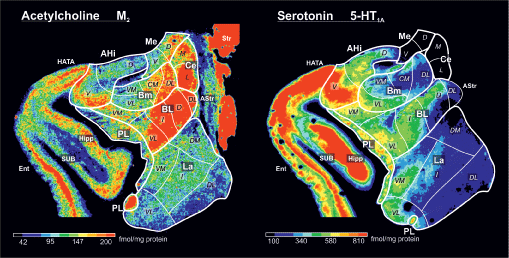
Figure 1: Distribution of Cholinergic M2 and Serotoninergic 5-HT1A Receptors in the Human Amygdala.
Subdivisions of the amygdala were identified in the color-coded receptor autoradiographs immediately adjacent to Nissl-stained sections in the right hemisphere at the level of the central nucleus. Color scales visualize the concentrations for each receptor (cholinergic muscarinic M2 or serotoninergic 5-HT1A receptors). After incubation of 20-μm thick coronal sections with the respective tritium-labelled ligand and exposition of the sections against the tritium-sensitive films, the films were developed, digitized and transformed into the binding site concentrations (fmol/mg protein) by using a nonlinear calibration curve between co-exposed plastic [3H]-standards of a known, step-wise increasing radioactivity and the gray values of the actual autoradiograph [1].
Amygdaloid subdivisions (mapping is based on the parcellation scheme of the previous study [2]: AHi - amygdalohippocampal transition area (from the superficial amygdaloid group), Bm - basomedial nucleus, BL - basolateral nucleus, La - lateral nucleus, PL - paralaminar nucleus (constitute the laterobasal group), Me - medial nucleus, Ce - central nucleus (constituents of the centromedial group, classification from [2]).
Parts of the subdivisions: CM - caudomedial, D - dorsal, DL - dorsolateral, DM - dorsomedial, L - lateral, M - medial, I - intermediate, V - ventral, VL - ventrolateral, VM - ventromedial. Adjacent structures: AStr - amygdalostriatal transition area, Str - striatum, HATA - hippocampal-amygdaloid transition area, Hipp - hippocampus, SUB - subicular complex, Ent - entorhinal cortex.