Meyerson phenomenon (MP) is an uncommon clinical condition that is characterized by an eczematous halo surrounding a preexisting melanocytic nevus (MN) and numerous other lesions. The etiology is unknown and the main concern is malignant transformation. We report an original case of a MP simulating a halo nevus.
Meyerson phenomenon, halo nevus, malignant transformation, histopathological examination
MP: Meyerson Phenomenon; MN: Melanocytic Nevus; MyN: Meyerson Nevus
MP is an uncommon clinical condition that is characterized by an eczematous halo surrounding a preexisting MN and numerous other lesions [1]. The etiology of this condition is unknown and the main concern with it is malignant transformation [2]; hence the interest of our case report.
We report a case of a 40-year-old woman, with no significant past medical history. Who presented with a cutaneous lesion which existed for her whole life. In the past 2 months, it has become scaly and itchy from no apparent cause. Clinical examination showed a 15 mm brownish nodule, situated on the lower back, surrounded by an irregular hypopigmented and lightly reddish halo topped by scales (Figure 1).
Treatment was surgical excision of the lesion with 2 mm margins. Histopathological examination revealed a dermal nevus, with epidermal hyperkeratosis, vesicles, spongiosis and perivascular lymphocytic infiltrate around it. Based on the clinical and histopathological findings, a diagnosis of Meyerson nevus (MyR) was made.
After treatment, there was complete resolution of the eczematous eruption and the hypopigmented halo (Figure 2). No recurrence was observed during 9-month follow-up.
MP, also termed MyN or halo dermatitis, is an inflammatory reaction surrounding a preexisting MN and numerous other lesions [1]. Meyerson first described this rare finding in 1971 [3].
This condition is typically seen in the trunk and proximal extremities of healthy young adults, with male predominance. A preexisting nevus may present with pruritus and scaling over the lesion. The eczematous halo is sharply defined and surrounds a central nevus symmetrically. In two-thirds of cases, multiple nevi are involved and can present either separately over time or simultaneously [2].
Common histological features, which were consistent with our patient, are spongiosis with a cellular infiltrate made of lymphocytes and eosinophils. Other characteristics include irregular acanthosis, parakeratosis and an unchanged nevus [4].
However, MP is not limited to benign MN. It can be present in atypical nevi and non-melanocytic lesions such as seborrheic keratosis, molluscum contagiosum, dermatofibromas, stucco keratosis, lentigo, keloid, and insect bites, as well as basal cell and squamous cell carcinomas [3].
The pathogenesis of MP is unknown. It has been suggested that it could be the result of allergic contact dermatitis, a hypersensitivity reaction, or a response to solar exposure, or some medications. There is substantial evidence that halo dermatitis is immune-mediated, by CD4 T lymphocytes as the major cellular infiltrate [3,5].
MyN can be mistaken for malignant melanoma or Sutton's nevus, also known as halo nevus. The latter begins as a benign nevus which evolves toward a zone of depigmentation followed by regression of the nevus. Histopathology will help in the differential diagnosis showing a dense inflammatory infiltrate mainly made by CD8 T lymphocytes [5,6].
The concern with benign lesions presenting with inflammation is malignant transformation [3].
The dermatitis can resolve with excision or spontaneously within a few months without any involution of the nevi. It has also been shown to clear with application of potent topical steroids [3].
MP is rarely mentioned in the dermatology literature. The main concern with this condition is malignant transformation; thus, clinicians need to be more aware and consider it in the differential diagnosis of itchy melanocytic lesions.
None.
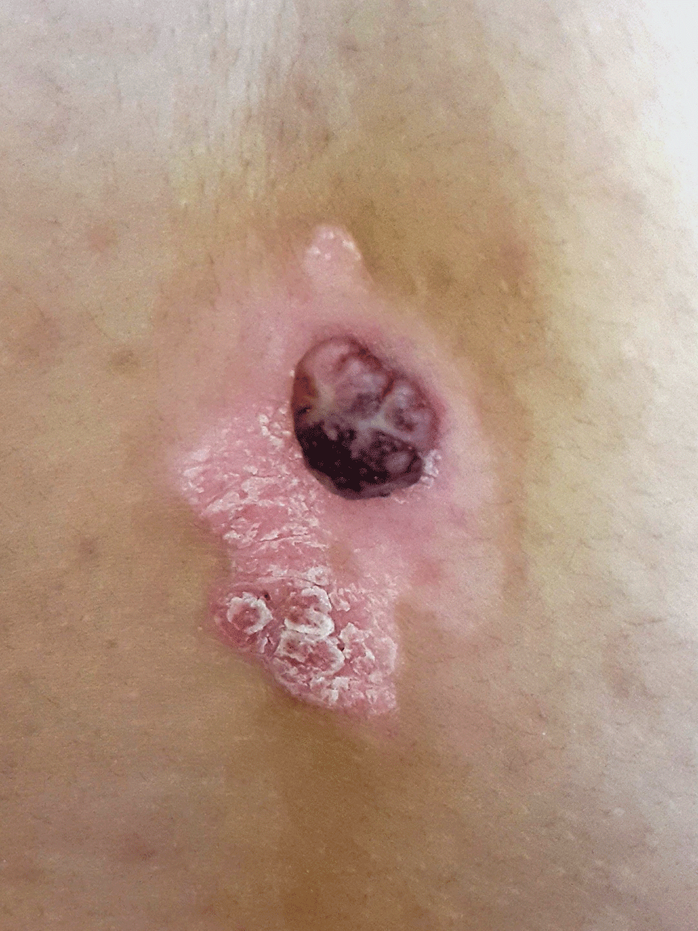
Figure 1: 15 mm brownish nodule, situated on the lower back, surrounded by an irregular hypopigmented and lightly reddish halo topped by scales.
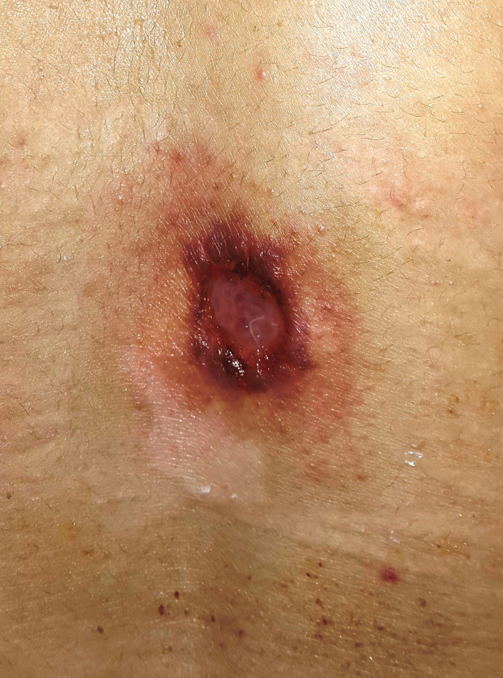
Figure 2: Control photo one month after surgery, showing the resolution of the eczematous eruption and the hypopigmented halo.