A 68-year-old man with a known history of cholelithiasis presented with worsening epigastric and right upper quadrant discomfort. He was afebrile, with mild leukocytosis (13.3 K/CMM) and normal liver function tests. Ultrasound and CT (abdomen/pelvis) showed cholelithiasis with gallbladder wall calcifications (Figure 1A, Figure 1B and Figure 1C, arrow indicates calcifications) and an incidental 7 cm right renal mass. Patient underwent laparoscopic cholecystectomy in conjunction with laparoscopic radical nephrectomy. Intraoperative findings demonstrated porcelain gallbladder with dense fibrotic adhesions. Pathology of the gallbladder revealed cholelithiasis, chronic cholecystitis with focal high-grade dysplasia and extensive calcified fibrosis of gallbladder wall. Right kidney pathology showed 5.4 cm clear cell renal cell carcinoma (WHO/ISUP Grade 2) with clear margins. The patient had an uneventful recovery. Although historically associated with increased risks of malignancy, the true incidence of malignancy in porcelain gallbladder is thought to be significantly lower than previously reported. Operative management is indicated in symptomatic patients.
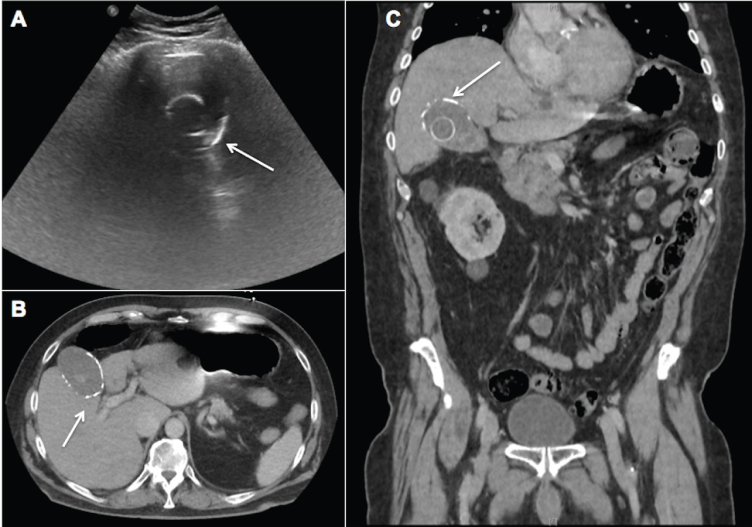
Figure 1: Ultrasound of the abdomen (A) demonstrates calcification in the wall of the gallbladder (arrow); which is also documented on CT scan (B & C, arrow). A calcified gallstone is also noted on ultrasound (A) and CT scan (C).