A 33-year-old female was admitted to COVID ICU after testing positive for the disease with 7 days history of fever, cough, inability to open as well as pain in the left eye, and inability to speak with right sided hemiparesis. She was known diabetic and hypertensive. Clinical examination showed presence of multiple pus points in left infratemopral region. Its microscopic examination showed wide area of necrosis along with cluster of mucormycosis. Due to sudden deterioration of patients GCS, CECT head was done (Figure 1) which showed multiple irregular thick walled peripherally enhancing lesion involving the left frontal, parietal and basal ganglia. It also showed presence of significant perilesional edema causing mass effect and midline shift (Figure 1). Patient underwent emergency debridement and burr hole drainage of abscess along with placement of external ventricular drain (Figure 2). The possible mechanism could be that the fungus has gained access to the brain from orbit or paranasal sinus resulting in sequential involvement and abscess formation. Gradually, patient condition deteriorated with progression of disease and she died due to refractory shock and extensive brain involvement.
None.

Figure 1: CECT head showing multiple irregular thick walled (green) peripherally enhancing lesion (red arrow) along with fronto-temporo-parietal infarct (blue arrow) with mass effect and midline shift.
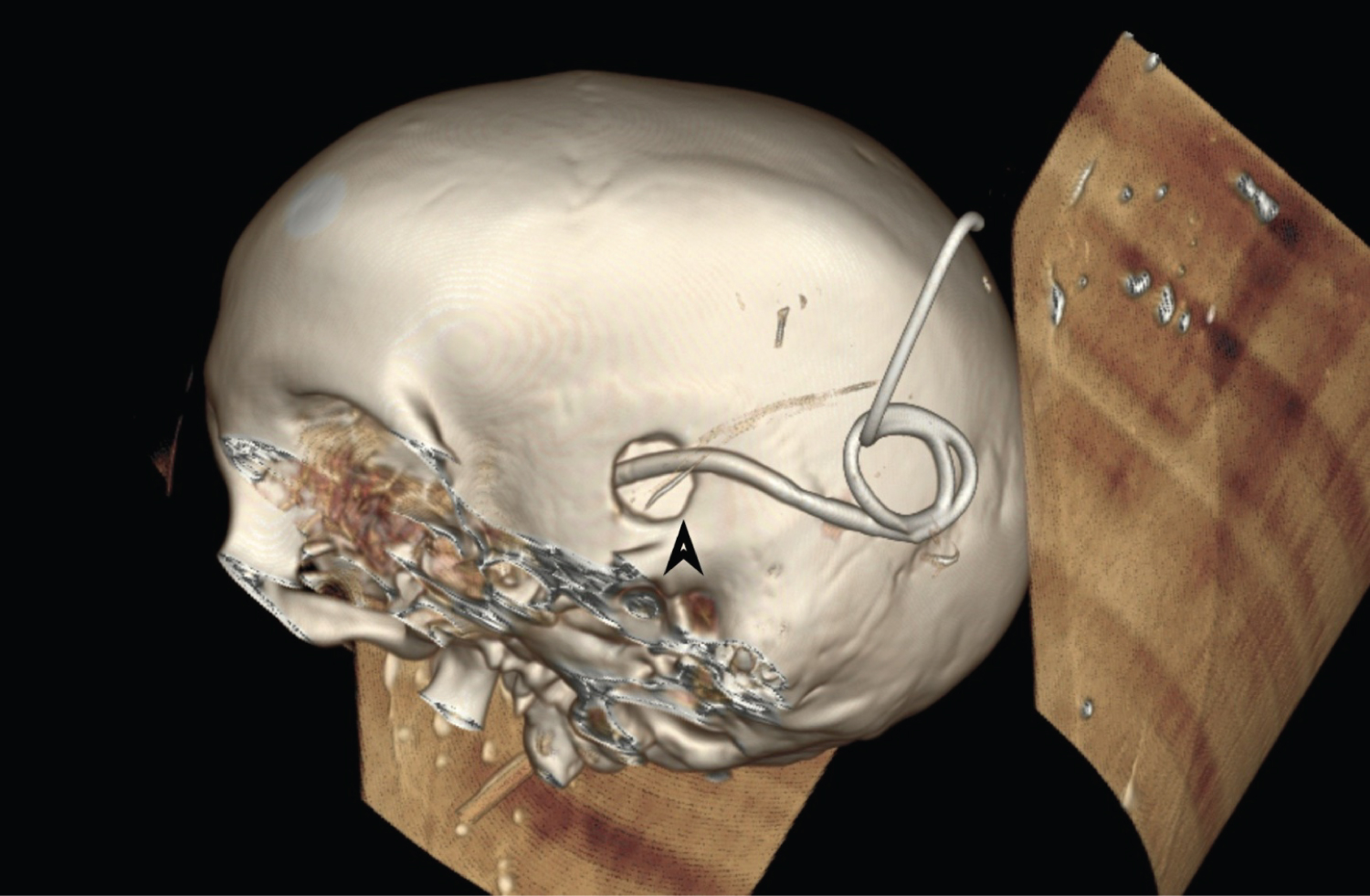
Figure 2: 3D representation of Skull showing burr hole and external ventricular drain in situ (black arrow).