Emphysematous pyelonephritis is a necrotizing infection of the renal parenchyma and surrounding tissues, characterized by the presence of gas. It is a serious infection whose main complications are septic shock, acute renal failure and disseminated intravascular coagulation [1]. Subcapsular haematoma secondary to pyelonephritis is an extremely rare complication whose clinical manifestations vary considerably according to terrain and severity [2].
We report the case of a 41-year-old female patient, a known diabetic on insulin, who presented to the emergency department with left lumbar pain evolving for 3 days in a febrile context. She also reported irritative urinary signs associated with cloudy urine. On admission, performance status was 2, the patient was conscious with a temperature of 38.9 ℃ and significant pain on palpation in the left lumbar fossa. Emergency blood tests revealed an inflammatory syndrome and impaired renal function. Standard radiography revealed a stone in the left renal area. CT scan (Figure 1) revealed an enlarged kidney with pyelocalic dilatation upstream of a pyloric calculus, and a heterodense subcapsular perinephric collection of hematic density and multiple air bubbles. This collection measures 6 cm thick medially. There is also extensive infiltration of peri-renal fat. Emergency treatment consisted of a venous line and antibiotic therapy with ceftriaxone and amikacin, adapted to her renal function. She was rushed to the OR for a double-J catheter (purulent urine) and percutaneous nephrostomy. The final outcome was favorable, with improvement of clinical signs and normalization of biological parameters.
Diagnosis of emphysematous pyelonephritis and subcapsular hematoma of the kidney is based on imaging [3]. Management is medico-surgical, involving bypass/drainage (double J catheter, percutaneous nephrostomy) or nephrectomy [1].
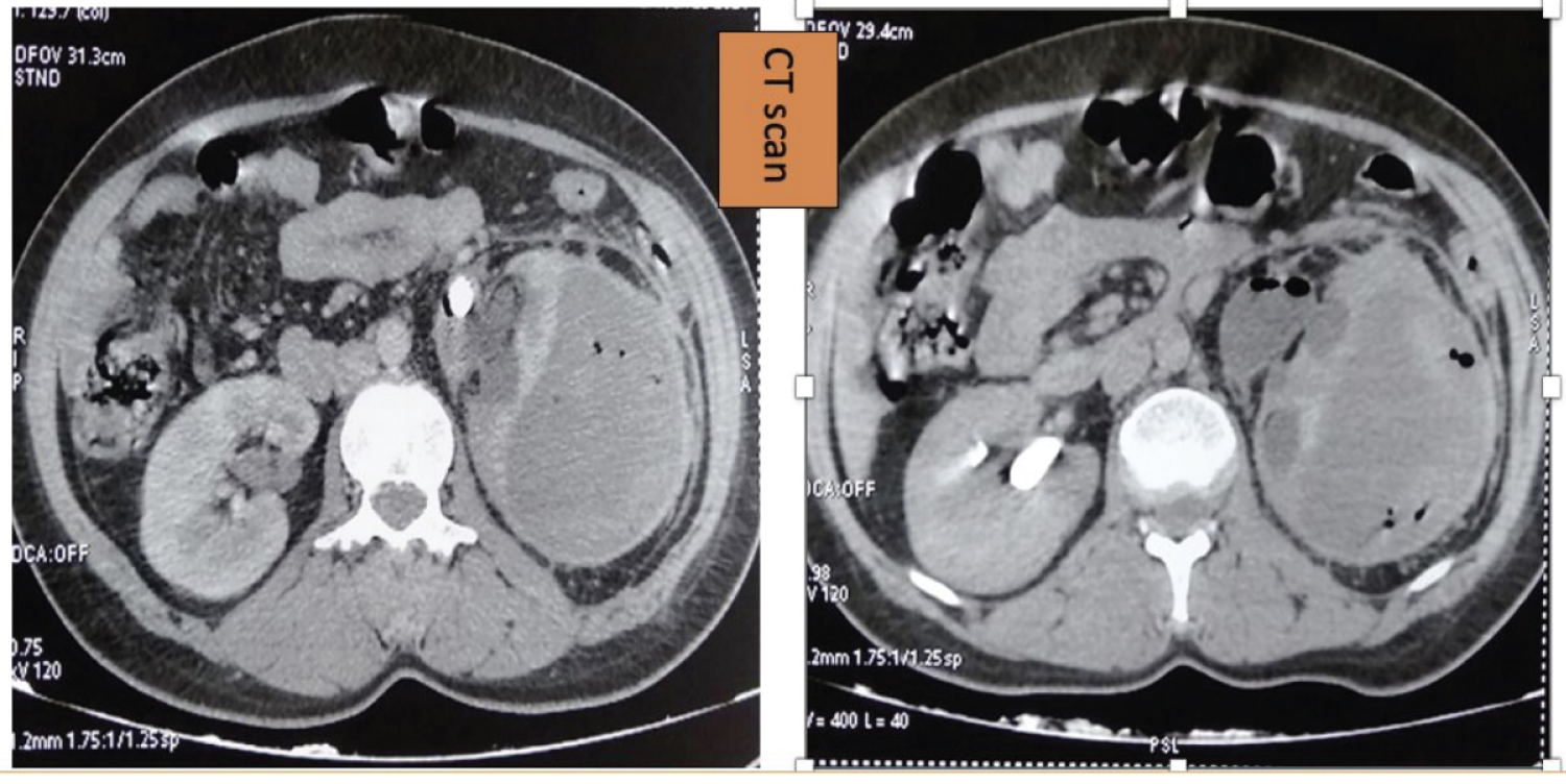
Figure 1: CT sections showing a hypertrophied kidney with pyelo-caval dilatation upstream of a pyloric calculus, and a heterodense subcapsular perinephric collection with patches of hematic density and multiple air bubbles.