

International Archives of Urology and Complications
Late Complications of Duplex System Ureterocele; Acute Urinary Retention, Stone Formation and Renal Atrophy
Sipal Timucin1* Akdere Hakan2 and Bumin Ors1
1Department of Urology, Cerkezkoy State Hospital, Turkey
2Trakya University Health Center for Medical Research and Practice, Turkey
*Corresponding author:
Sipal Timucin, Department of Urology, Cerkezkoy State Hospital, Tekirdag, Turkey, Tel: +905548430218, E-mail: drtimucinsipal@gmail.com
Int Arch Urol Complic, IAUC-1-008, (Volume 1, Issue 2), Case Report; ISSN: 2469-5742
Received: June 28, 2015 | Accepted: August 10, 2015 | Published: August 14, 2015
Citation: Timucin S, Hakan A (2015) Late Complications of Duplex System Ureterocele; Acute Urinary Retention, Stone Formation and Renal Atrophy. Int Arch Urol Complic 1:008. 10.23937/2469-5742/1510008
Copyright: © 2015 Timucin S, et al. This is an open-access article distributed under the terms of the Creative Commons Attribution License, which permits unrestricted use, distribution, and reproduction in any medium, provided the original author and source are credited.
Abstract
A 49- year-old woman was admitted to emergency department with a complaint of acute urinary retention. The investigation of the patient revealed right duplex system anomaly, ureterocele containing multiple stones and atrophic right kidney. After reliefing her urinary retention, endoscopic ureterocele de-roofing, two dj stents insertion and stones extraction were performed. The symptoms of the patient were relieved after treatment. The patient was asymptomatic at six month follow-up visit.
Keywords
Duplex system ureter, Multiple calculi, Transurethral ureterocele incision, Ureterocele, Renal atrophy
Introduction
Ureterocele is cystic dilation of the terminal ureter and its incidence among newborns was reported 1/500 - 1/4000 [1]. It may be associated with tissue defect of bladder, bladder neck and posterior urethra. Eighty percent of ureteroceles are seen in the ureter draining the upper pole of a complete ureteral duplication. The cases whose diagnoses are omitted in early ages may suffer from recurrent urinary tract infection, stone formation, septicaemia and renal failure in later years. They generally break out in single system, orthotropic and intravesical in adults [2]. Ureteral atony and urinary stasis in ureterocele may result in stone formation. The incidence of stone in ureterocele was reported between 4%-39% and it varies depending on patient's geographic origin and occurs generally in male patients [3]. Endoscopic ureterocele incision and stone extraction was recommended as an effective treatment in adults with minimal risk of postoperative reflux [4]. Although the stone formation in ureterocele has been mentioned in many reports, the late presented case with acute urinary retention and stone containing ureterocele in unilateral atrophic duplex system is very rare.
Case
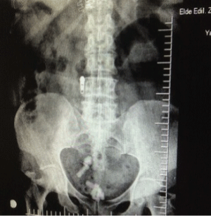
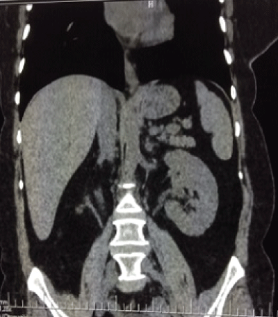
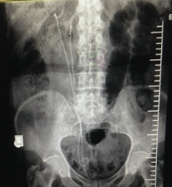
A 49-year- old woman was admitted to emergency service with acute urinary retention due to an obstructed urethral stone. After relieving retention with the insertion of urethral catheter, direct kidney urether and bladder (KUB) x-ray and urinary ultrasonography (USG) were performed. KUB x-ray revealed multiple opacities at right lower quadrant of minor pelvis (Figure 1). Her medical history revealed that she had experience recurrent square shaped urinary stone pass and hadn't had any urological intervention. Ultrasonography demonstrated a right side terminal ureteral cystic dilatation, containing multiple stones and atrophic duplex systems (Renal bipolar length: 5cm, renal parenchymal thicknesses 0, 5cm, and dilatation of the lower system pole). For further investigation a non-contrast tomography showed right terminal ureteral dilatation (3×2×2cm), multiple calculus in 1×1cm size and duplex system anomaly (Figure 1-3).

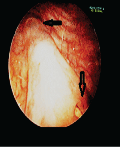
Renal Dimercaptosuccinic acid (DMSA) scintigraphy were performed and showed 7% split right renal function. Her urine culture was clear and the pH was 6.0, Blood Urea Nitrogen (BUN): 45mg/dl, creatinine: 1.4mg/dl. After receiving the informed consent form the patient underwent cystoscopy and transurethral ureterocele de-roofing surgery (Figure 4,5).
.
Figure 4: Arrows at cystoscopy showed upper pole orifice and a stone in the lower pole ureterocele orifice
View Figure 4
.
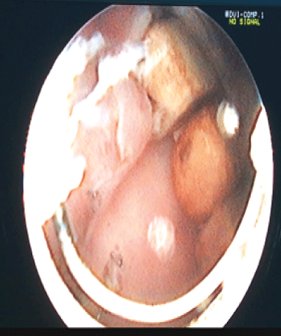
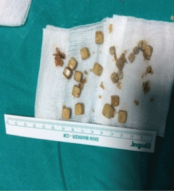
Figure 5: Transurethral ureterocele deroofing was performed and cube shaped stones were revealed
View Figure 5
Her stones were removed from the ureterocele followed by double J (DJ) stents were inserted (Figure 6,7). The control x-ray confirmed stone-free status postoperatively. The result of stone analysis revealed that the 60% of compound was carbonate apatite and 40% of it was calcium oxalate. DJ stents were removed after 1 month. She was asymptomatic at 6 months follow- up visit.
Discussion
Ureteral duplication with 1% incidence of population is one of the most common congenital urologic anomalies. 70% of the cases are female and 20% are bilateral [4]. It's more possible to see congenital anomalies in later adulthood in rural areas or they can be found out by chance. According to the literature, the incidens of stones in ureteroceles lies between 4-39% and most stones solitary [3] .Ureteroceles including stones tents to cause symptoms such as Urinary tract infections (UTI), bladder irritability and flank pain. Most duplex system ureteroceles present with urinary tract infections at an early age, if not recognized before birth by ultrasound and adult presentation is uncommon. Approximately twenty percent of the duplex systems have complete double ureters which open to different localisations in lower urinary tract. Complete duplex ureters usually comply with Weigert-Meyers rule, the upper pole moiety has ectopic insertion medial and inferior to the lower pole moiety ureter and more prone to obstruction [2]. Ureteroceles in asymptomatic adults usually occur in a single system and stone containing ureteroceles are usually seen in males [3]. Unlikely the other previously reported cases, our patient was female and had a complete duplication of right collecting system and lower moiety ending in uroterocele obstructed by stones. It is not necessary to perform open surgery contrary to the treatment in the pediatric age group. Endoscopic incision and stone extraction have been reported as an effective treatment in adults with minimal risk of reflux [4]. The patient underwent transurethral ureterocele de-roofing and stones were removed from the ureterocele. Her symptoms were relieved and she was asymptomatic at six months follow- up.
This case is important not only due to its rarity but also it underscores the importance of timely imagine studies in patient with recurrent stone pass history and it revealed that delayed diagnosis ureteroceles may be presented with serious late complications.
References
-
Campbell M (1951) Ureterocele; a study of 94 instances in 80 infants and children. Surg Gynecol Obstet 93: 705-718.
-
Schlussel RN, Retik AB (2007) Ectopic ureter, ureterocele and other 5. Anomalies of the ureter. In: Wein AJ, Kavoussi LR, Novick AC, et al. (eds). Campbell-Walsh urology. 9th edn. Philadelphia: Saunders, 3383-3422.
-
Messing EM, Henry SC (1979) Stones in orthotopic, non-obstructing ureteroceles. J Urol 122: 403-404.
-
Shah HN, Sodha H, Khandkar AA, Kharodawala S, Hegde SS, et al. (2008) Endoscopic management of adult orthotopic ureterocele and associated calculi with holmium laser: experience with 16 patients over 4 years and review of literature. J Endourol 22: 489-496.
