
Trauma Cases and Reviews
Analysis of a Clinically Failed, Mechanically Intact, Reconstructive Compression Plate
Bharadwaj Cheruvu1, Sunil Karmacharya1, Richard Laughlin2 and Tarun Goswami1,2,3*
1Department of Biomedical, Industrial and Human Factors Engineering, Wright State University, USA
2Orthopaedic Surgery, Sports Medicine and Rehabilitation, Wright State University, USA
3Institute of Materials Science and Welding, Graz University of Technology, Austria
*Corresponding author: Tarun Goswami, Department of Biomedical, Industrial and Human Factors Engineering, Orthopaedic Surgery, Sports Medicine and Rehabilitation, Wright State University, 3640 Colonel Glenn Hwy Dayton, OH 45435, USA, Tel: 937-775-5120, Fax: 937-775 -7364, E-mail: tarun.goswami@wright.edu
Trauma Cases Rev, TCR-1-007, (Volume 1, Issue 2), Research Article; ISSN: 2469-5777
Received: June 24, 2015 | Accepted: September 05, 2015 | Published: September 07, 2015
Citation: Cheruvu B, Karmacharya S, Laughlin R, Goswami T (2015) Analysis of a Clinically Failed, Mechanically Intact, Reconstructive Compression Plate. Trauma Cases Rev 1:007. 10.23937/2469-5777/1510007
Copyright: © 2015 Cheruvu B, et al. This is an open-access article distributed under the terms of the Creative Commons Attribution License, which permits unrestricted use, distribution, and reproduction in any medium, provided the original author and source are credited.
Abstract
A reconstructive orthopedic bone plate was submitted for analysis. Traditional failure analysis methods were used to assess the mode of the plate failure. Metallographic investigation of the plate was carried out in this report. Since limited data was available in the literature and clinical data related to subject demography, date of removal as well as reasons for removal unknown, in depth analysis was not possible. However, the plate was received in in-tact condition with minor biological deposits and scratches, it is speculated that the failure of the plate may have been due to biological/clinical reasons, likely infection, rejecting the device due to lack of union at the fracture site.
Keywords
Failure analysis, compression plate, locking screws, fracture, corrosion, scratches
Introduction
Proximal femoral fractures are quite commonly seen among orthopedic physicians. Between 220,000 and 250,000 proximal femoral fractures occur in the United States each year [1]; 90% of these fractures occur in patients older than 50 years [1]. In younger patients, proximal femoral fractures are usually the result of high-energy physical trauma (e.g., high-speed motor vehicle accidents) and usually occur in the absence of disease. Intertrochanteric and femoral neck fractures account for 90% of the proximal femoral fractures occurring in elderly patients [2]. Proximal femoral fractures in elderly patients are often pathologic, usually resulting from minimal-to-moderate physical trauma to areas of bone significantly affected by osteoporosis.
Majority of fractures among the elderly are often due to a sideways fall from a standing height [2]. This mechanism can be explained by a displacement of the greater trochanter.
Fixation devices for intertrochanteric fractures include intramedullary devices (e.g., Gamma nail, intramedullary hip screws, etc.) and extramedullary devices, mainly sliding screws and plates (e.g., dynamic hip screws). The former showed an advantage in fixation of unstable intertrochanteric fractures and the latter yielded better results for fixation of stable intertrochanteric fractures [3]. The correct insertion and positioning of both intra- and extramedullary devices are known to prevent implant failure and cutout.
The primary benefits from plate include: 1. Increase fracture healing rate, 2. Decrease loss of bone mass shielded by plate and 3. Decrease incidence of refracture of bone after removal of plate. The weakest region in the plate can be found in the screw hole as a result of increased stresses. A more flexible plate allows for micromotion and restoration of blood supply to the bone. The most common failure modes of a bone-plate-screw fixation are screw loosening and plate failure. Plate failures are often caused by fatigue or crevice corrosion through the screw hole [4].
Compression plates are widely used because of the need for compression between the fracture fragments. The advantages of these compression plates include a lower incidence of malunion, stable internal fixation, and movement of neighboring joints due to lack of use of external fixators. The advantages and success of these compression plates are due to surgical technique. However, the plating system has some disadvantages. Those disadvantages include a delayed union as well as a persistence of a microscopically detectable fracture gap which can act as a stress riser after plate removal. In addition, cortical bone loss under the plate can also be evident as a disadvantage.
Cortical bone loss can be explained from the mechanism in which the unloading of bone leads to loss in bone, often induced by stress shielding. Bone loss as a result from stress shielding, is often a direct result of a resorptive process defined by porosis of the endosteal half of the cortex during the early phases of plate fixation and culminating in the loss of porotic endosteal bone during the late phases. Due to loss of endosteal bone, the medullary canal widens and the cortices narrow.
Plates and screws are widely used in bone fixation and in comminuted fractures which have load-bearing fixation applied across the area of comminution. A reconstructive plate with at least three and preferably four screws on each side of the comminuted area bear optimal load at fixation. As the treatment of fracture using reconstructive plate increase, more clinical outcome data would be available in the future [5]. To date, there are no studies which have investigated the failure of these devices. This study was therefore undertaken to experimentally evaluate the surgically failed, mechanically intact, reconstructive plate made of stainless steel 316L which was fixed to the bone at the site of the fracture. The reasons for removal can be due to either mechanical or biological causes.
Materials and Methods
Reconstructive locking plate
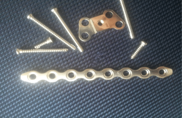
The submitted reconstructive plate was a 8 hole, 105.5 mm long, 2.8mm thick along with 2 locking head screws (4.3 mm diameter and 14 mm long) and 4 non-locking screws (4.3 mm diameter and 37 mm long) were submitted for the failure analysis. Both plates and locking head screws are made per specification ASTM 240 and ASTM 666 [6]. The plate was labeled properly with a serial number (Figure 1). Even though the subject demography, implant site, date of removal and reason for removal was unknown, the analysis of the plate was carried out with visual, optical, and scanning electron microspores and surface topography mapped.
Visual examination of the plate
Macroscopic inspection of plate and screws revealed scratches on the surface of the plate along with some debris particles confirmed that the plate was prematurely removed from the fracture site. Both locking screw and non-locking screws were examined visually and no significant damage was found on the screws.
Analysis using optical microscopy
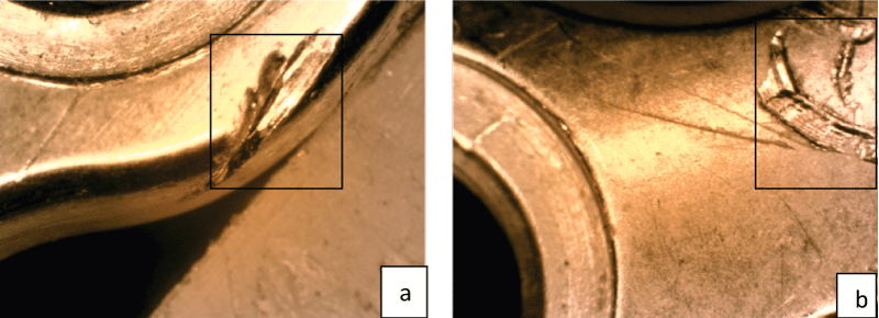
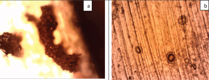
The plate contained scratch marks as well as some corrosion and debris on the surface and holes see Figure 2 and Figure 3.
.
Figure 2a and 2b: Marked surface of the plate with scratches and pits (mag: 10X)Marked surface of the plate with scratches and pits (mag: 10X)
View Figure 2a and 2b
.
Figure 3a: Debris in the surface of plate in 200X mag
Figure 3b: surface with pitting
View Figure 3a and 3b
Hardness of material
The average hardness of the plate was found to be 286 HB (Brinell hardness) that corresponds to a tensile strength of 965 MPa. These are slightly higher than the specifications.
Results and Discussion
The plate was received in the intact form. Since the plate was used in vivo some damage due to exposure was documented in this paper in the form of corrosion pits, debris (biological in nature) and scratches, though it is unknown whether those scratches occurred during exposure, removal or transportation and storing of the device. The material has met the specification requirements and thus was a successful design per engineering considerations.
This case is unique in that two cultures with different expectations were attempted to be bridged. While the plate maintained the engineering integrity, it failed in surgery because of the following likelihood:
1) Removed upon healing occurred to the satisfaction of the patient and surgeon, however, it is known that revision surgery took place once this device was removed from the fracture site.
2) Biological factors and reactionary forces did not accept the device and those scenarios may result in a lack of union of the fractured bones, infection due to metal particles, and improper placement of the device, resulting in removal and revision.
Conclusion
The engineering failure analysis found the device to conform to the specifications and components maintained structural integrity, thus it was not a failure. Surgical outcome was compromised may be due to infection, requiring the device removal and revision surgery. Therefore, this example is a complex case, and must be considered in the design and development of orthopaedic medical devices.
References
-
Zuckerman JD (1996) Hip fracture. N Engl J Med 334: 1519-1525.
-
Helgason B, Gilchrist S, Ariza O, Chak JD, Zheng G, et al. (2014) Development of a balanced experimental-computational approach to understanding the mechanics of proximal femur fractures. Medical engineering & physics 36: 793-799.
-
Parker MJ, White A, Boyle A (2008) Fixation versus hemiarthroplasty for undisplaced intracapsular hip fractures. Injury 39: 791-795.
-
Uhthoff HK, Poitras P, Backman DS (2006) Internal plate fixation of fractures: short history and recent developments. J Orthop Sci 11: 118-126.
-
Thapa N, Prayson M, Goswami T (2015) A failure study of a locking compression plate implant. Case Studies in Engineering Failure Analysis 3: 68-72.
-
(2005) ASTM F86-04: Standard Practice for Surface Preparation and Marking of Metallic Surgical Implants, Annual Book of ASTM Standards 13.01.
