
Trauma Cases and Reviews
Fracture-Dislocation of the Upper Thoracic Spine without Neurologic Associated Injury: Two Cases Report
J Miguel Rodríguez Solera*, Ana María Cerván de la Haba, Miguel S Hirschfeld León and Enrique Guerado Parra
Department of Orthopaedic Surgery, Traumatology and Rehabilitation, Hospital Universitario Costa del Sol, Spain
*Corresponding author:
José Miguel Rodríguez Solera, Hospital Universitario Costa del Sol, Marbella, España, Autovía A7 km 187 C.P. 29603, Spain, Tel: 951976224, E-mail: jmyl09@icloud.com
Trauma Cases Rev, TCR-1-022, (Volume 1, Issue 4), Case Report; ISSN: 2469-5777
Received: October 30, 2015 | Accepted: December 19, 2015 | Published: December 22, 2015
Citation: Solera JMR, de la Haba AMC, León MSH, Parra EG (2015) Fracture-Dislocation of the Upper Thoracic Spine without Neurologic Associated Injury: Two Cases Report. Trauma Cases Rev 1:022. 10.23937/2469-5777/1510022
Copyright: © 2015 Solera JMR, et al. This is an open-access article distributed under the terms of the Creative Commons Attribution License, which permits unrestricted use, distribution, and reproduction in any medium, provided the original author and source are credited.
Abstract
Upper thoracic fractures are produced by high-energy trauma. Fracture-dislocation (type C Association) for the Study of Internal Fixation (AO classification) of the upper thoracic spine represents 15% of all thoracic and lumbar fractures. The neurological injury occurs in 80% of cases, being extremely rare type C fractures without spinal cord injury.
We report a case of 51 years-old male with no past medical history, who was brought to our Hospital after falling from a 10 meters high roof. He had 15 points at Glasgow Coma Scale (GCS) and a completely normal neurological examination. A whole body computerized tomography (CT) scan was performed and diagnosed a fracture-dislocation at T8-T9. Acute surgery was decided, performing an open reduction and internal fixation with pedicle screws from T6 to T1. Clinical outcome was favorable.
The second case is a 29 years old patient, who suffered a motorcycle accident. On arrival at critics room presented 14 points at GCS and American Spinal Injury Association scale (ASIA) E. After whole CT scan, was diagnosed a T10 fracture-dislocation, an open left iliac fracture, open right distal tibia fracture and a fracture of 10th left costal arch. Acute surgery was decided: performing external fixation of right tibia, wound cleaning and exploration of iliac crest and open reduction and fixation of the fracture-dislocation of thoracic spine. After nine days from the first surgery an open reduction and internal fixation (ORIF) of the right distal tibia was performed and a closed reduction and internal fixation (CRIF) of tibial plateau fracture. Ligamentoplasty of anterior cruciate ligament (ACL) of the right knee was also done arthroscopically. Clinical outcome was satisfactorily.
The fracture-dislocation of the upper thoracic spine is often associated with spinal cord injury, there is a very few reported cases in bibliography without neurological damage. The initial clinical management of this patients is crucial at critics room, Advanced Trauma Life Support (ATLS) must be applied and subsequently perform a damage control. In patients with upper thoracic fracture-dislocation without spinal cord involvement, immediate reduction and osteosynthesis should always be performed.
Keywords
Unstable thoracic fractures, Dislocation, Upper thoracic spinal fractures
Case Report
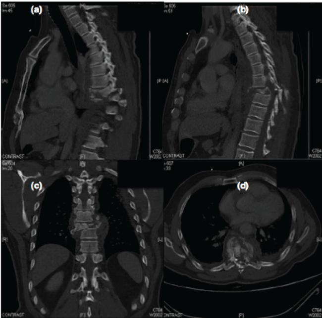
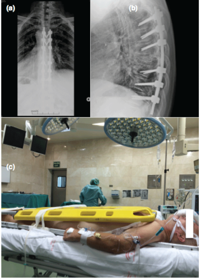
We report a case of 51 years-old male with no past medical history, who was brought to our Hospital after has fallen from a 10 meters high roof while working. Physical examination at time of arrival was: hemodynamically stable, 15 points at GCS, thoracic pain and scalp erosions. On neurological examination he presented hyperalgesia at T7, T8, T9 level (ASIA E). After clinical stabilization a whole body angio CT-scan was performed which revealed a spinous process fracture of T6, left articular process fracture of T7, fracture-dislocation of T8-T9 with burst fracture of T8 (AO classification: A0 T6, A0 T7, C T8-T9, A4 T8, A1 T9), bilateral ribs fractures, pneumothorax, pneumomediastinum and bilateral pleural effusion (Figure 1). Emergent (< 6 hours) open reduction and posterior instrumentation with transpedicular screws and two rods was performed (Figure 2). Fully improved after a year from surgery.
.
Figure 1: (a) Sagittal CT; (b) Sagittal CT; (c) Coronal CT; (d) Axial CT. CT images demonstrates a fractures at T6 (AO), T7 (AO), T8T9 (C), T(8) (A4) y T9 (A1) levels (AO classification).
View Figure 1
.
Figure 2: (a) Anteroposterior spine radiograph after spine surgery; (b) Lateral spine control radiograph; (c) Patients with this type of unstable lesson must be perfectly immobilized for the chance to a prone position, we used for that two shelves.
View Figure 2
The second case is a 29 year old patient with no medical history, which is transmitted by the emergency services to our center after a motorcycle accident. In emergency room, he presented acceptable overall, GCS 14 (sedated). Blood pressure 90/40 mmHg, free airway and normal breathing. Pulse preserved, rhythmic. Neurological examination without significant pathological findings only referred altered sensitivity in anterior left thigh. Presented a 30 cm left Ilioinguinal wound with iliac crest exposure, which is comminuted and with great involvement of soft tissues. He had also an open distal tibial fracture grade II (Gustilo and Anderson classification).
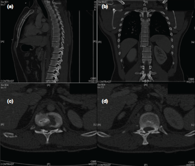
Ultrasound scan was performed without abdominal, pericardial or pleural fluids findings. After whole Body-CT scan was diagnosed of: Fracture-dislocation CA3 at T10 and A0 at T10 level (AO classification) (Figure 3), a 10th rib fracture, left iliac open fracture. Early surgical treatment is decided, performing Friedrich on left ilioinguinal wound, external fixation to the right tibia and open reduction and fixation with pedicle screws at T8-T12 (Figure 4). After 9 days from first surgery a second surgical procedure was performed by ORIF of right distal tibial fracture, right knee ligamentoplasty, ORIF of external tibial plateau fracture of right knee with two cannulated screws. The patient progressed satisfactorily nowadays, after three years of follow-up, no neurological injury is present.
.
Figure 3: Fracture disclocationat T10 level (a) Sagittal CT scan image; (b) Coronal CT scan image; (c and d) Axial CT scan images shows a dislocation of the left articular facet T10 vertebra.
View Figure 3
.
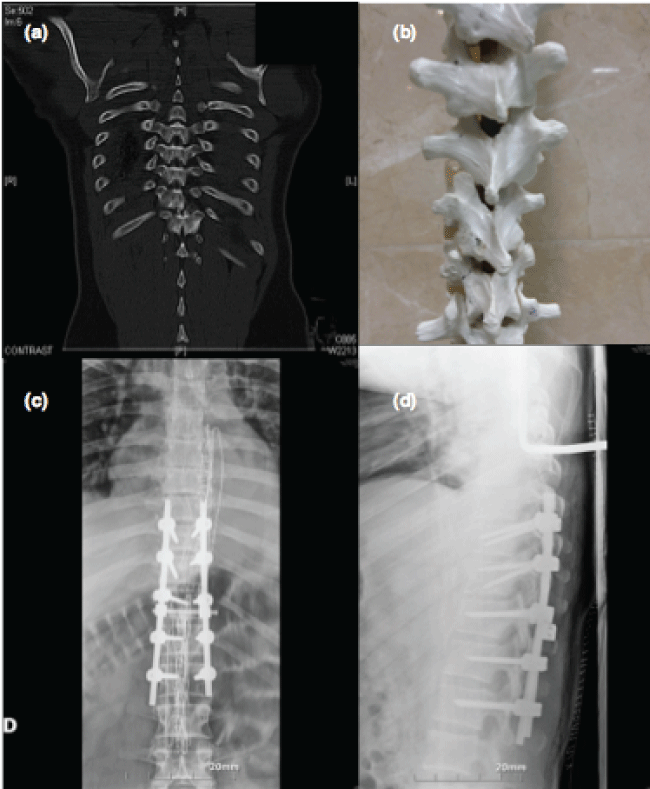
Figure 4: (a) Coronal CT scan image shows a fracture dislocation at T10 level; (b) Photograph demonstrate the injury at the plastic phantom; (c) Anteroposterior spine radiograph after surgery; and (d) Lateral spine radiograph illustrating the posterolateral instrumented fusion T8-T12.
View Figure 4
Discussion
Fracture-dislocation of the thoracic or lumbar spine caused by high-energy trauma often results in severe neurologic deficits when dislocation is significant. Patients are usually males, with an average age of 15-30 years old and traffic accidents are the most common cause. Account for 16 % of all vertebral fractures, up to 17 % of them are associated with another spinal fracture, associated thoracic injuries commonly and often-associated neurological injuries. Up to 83% of upper thoracic spine fractures have other injuries associated [1]. When a person falls from a height, the rib cage, the lungs, the heart and the liver present injury prevalence is greater than 50%. A fall from a height without suffering rib fracture is very infrequent and therefore questionable [2]. At the moment of impact, a falling body undergoes deceleration and the amount of kinetic energy transferred to the ground reacts with an equal amount against the body. The primary impact is usually the site of the most severe injury. In high falls, a compression fracture of the vertebrae is frequently observed [3].
Thoracic spine is divided into two regions: Upper thoracic spine (T1-T10) and thoracolumbar spine. Vertebral bones between T1-T10 form a kyphotic angle whose vertex is at T6 and T7, are stabilized by the rib cage, which increases by more than 30 % rigidity of thoracic spine [4,5].
The thoracic spine is much stiffer than the lumbar spine in sagittal and lateral flexion-extension, reflects the restraining effect of the rib cage, and the relatively thinner disks of the thoracic spine (average 20% of vertebral body height versus 40% in the lumbar spine) that restrict the arc of motion. Rotation about the craniocaudal axis, on the other hand, is greater in the thoracic spine, reaching its maximum at T8-T9. In the lumbar spine, rotation is limited by the orientation of the facets and the anterior portion of the annulus to only 10° for the entire lumbar spine, versus about 75° of rotation to each side in the thoracic spine [1].
Due to the relatively unstable thoracic spine of the fracture-dislocation, the spinal cord is extremely vulnerable due to in this region, the spinal canal is narrowed, with less free space between the cord and the osseous ring and the central thoracic spine also has a relatively sparse blood supply [1,6].
Management of patients with high thoracic spine fractures is one of the biggest challenges of the orthopedic surgeon.
Breakthroughs in the prehospital care enable patients arriving alive to the hospital for proper care.
The concept of damage control came up during the Second World War, initially for the management of abdominal injuries. Damage control surgery in orthopedics (CDO) is an extension of this concept.
The CDO has three steps: first (< 24 hours) early stabilization of unstable fractures and bleeding control, second improving the patient's condition in the intensive care unit and third (3rd-10th day) Final fracture stabilization in the best condition of the patient. In the case of isolated fractures of the spine, the treatment regimen is properly defined, however, the right time and the type of fixing to use in unstable thoracic and lumbar fractures in trauma patients remains controversial [7].
At the emergency room Advanced Trauma Life Support (ATLS) protocol should be follow (Airway, Breathing and ventilation, Circulation, Disability, Exposure and Environment). Regarding the image base diagnosis chest and pelvis anteroposterior (AP) and lateral cervical spine conventional radiography should be performed at the critics room and if the patient is hemodynamically stable, a whole body CT scan must be performed. In case of hemodynamic instability, ultrasound is recommended in the same critics room in order to dismiss abdominal, pleural or pericardial free fluid [8].
Once stabilized and diagnosed the injuries of patients, treatment consist in damage control protocol, first treating injuries in the head and chest or abdominal cavities, second stabilizing longs bones and pelvis and third, stabilization of the spine . In some instances, such as incomplete neurological injury progression, stabilization of the spine would have priority over the stabilization of long bones.
Timing management is a key role in these patients in order to avoid the second hit. Immediate surgery is recommended when there is neurological injury, spinal instability or dislocation.
Some authors advocate surgery in the first 6 hours after trauma and others in the first 24 hours, since it reduces stress and pain, and patient mobilization is not limited in the intensive care unit in addition to prevent secondary neurological impairment, thus avoiding aggravate lethal triad (acidosis, coagulopathy, hypothermia) [8].
The use of "spine damage control" fixation for unstable spine fractures is an evolving phenomenon in the management of patients with polytrauma. It consists of early stabilization of the unstable spinal injury with minimal surgical insult, followed by elective surgical stabilization and fusion [9].
The immediate availability of spine surgical equipment, the possible need for a second surgery or neurological decompression due to ligamentotaxis insufficient are some of the drawbacks of stabilization and fixation. In a second stage (window of opportunity) another surgery might be required (anterior approach) [8].
Conclusion
Fracture dislocations of the upper thoracic spine without neurologic deficit are very rare. Only few cases are reported [10-26]. It is important to apply ATLS protocol at the emergency room, and immediate surgery by reducing malalignment and fixation of spine. A second anterior approach must be assessed.
Conflicts of Interest
All authors have no financial support or relationships that may pose a conflict of interest to disclose for this work.
References
-
Luiz Roberto Vialle, Emiliano Vialle (2005) Thoracic spine fractures Injury. Injury Int. J. Care Injured 36: S65-S72.
-
Casali Michelangelo Bruno, Battistini Alessio, Blandino Alberto (2014) The injury pattern in fatal suicidal falls from a height: An examination of 307 cases. Forensic Sci Int 249: 52.
-
Vladimir Zivkovic, Slobodan Nikolic, Dragan Babic (2011) Pontomedullary Lacerations in Falls from a Height-A Retrospective Autopsy Study. Journal of Forensic Sciences 57: 654-657.
-
Morita D, Yukawa Y, Nakashima H, Ito K, Yoshida G, et al. (2014) Range of motion of thoracic spine in sagittal plane. Eur Spine J 23: 673-678.
-
Mannen, Erin, Anderson, John, Arnold, Paul (2015) Integrity of damage control posterior spinal fusion constructs for polytrauma patients: A biomechanical investigation. Spine 40: E760-E766.
-
Anthes TB, Muangman N, Bulger E, Eric JS (2012) Upper Thoracic Spine Fracture Dislocation in a Motorcyclist. Current Problems in Diagnostic Radiology 41: 128-129.
-
Stahel PF, VanderHeiden T, Flierl MA, Matava B, Gerhardt D, et al. (2013) The impact of a standardized "spine damage-control" protocol for unstable thoracic and lumbar spine fractures in severely injured patients: A prospective cohort study. J Trauma Acute Care Surg 74: 590-596.
-
Schmidt OI, Gahr RH, Gosse A, Heyde CE (2009) ATLS(R) and damage control in spine trauma. World J Emerg Surg 4: 9.
-
Pekmezci M, Herfat S, Theologis AA, Viscogliosi P, Demirkiran G, et al. (2015) Integrity of Damage Control Posterior Spinal Fusion Constructs for Patients With Polytrauma: A Biomechanical Investigation. Spine 40: E1219-E1225.
-
Enishi T, Katoh S, Sogo T (2014) Surgical Treatment for Significant Fracture-dislocation of the Thoracic or Lumbar Spine without Neurologic Deficit: A Case Series. J orthop case report 4: 43-45.
-
Gertzbein SD, Offierski C (1979) Complete fracture-dislocation of the thoracic spine without spinal cord injury. A case report. J Bone Joint Surg Am 61: 449-451.
-
Vichard P, de la Salle R, Tropet Y, Runge M (1983) Complete fracture-dislocation of thoracic vertebrae 8 and 9 without neurological complications. Description of the injury. Therapeutic deductions. Rev Chir Orthop 69: 645-648.
-
Weber S, Sutherland G (1986) An unusual rotational fracture-dislocation of the thoracic spine without neurologic sequelae internally fixed with a combined anterior and posterior approach. J Trauma 26: 474-479.
-
Harryman DT (1986) Complete fracture-dislocation of the thoracic spine associated with spontaneous neurologic decompression. A case report. Clin Orthop Relat Res: 64-69.
-
Sasson A, Mozes G (1987) Complete fracture-dislocation of the thoracic spine without neurologic deficit. A case report. Spine (Phila Pa 1976) 12: 67-70.
-
Uriarte E, Elguezabal B, Tovio R (1987) Fracture-dislocation of the thoracic spine without neurologic lesion. Clin Orthop Relat Res: 261-265.
-
Simpson AH, Williamson DM, Golding SJ, Houghton GR (1990) Thoracic spine translocation without cord injury. J Bone Joint Surg Br 72: 80-83.
-
Krallis P, Psicharis P, Dendrinos G (1992) Fracture-dislocation of the dorsal spine with severe displacement without neurologic impairment: Case presentation and literature review. Acta Orthop Belg 58: 84-87.
-
Miyasaka Y, Satomi K, Sugihara S, Tahara Y, Hayashi T, et al. (1993) Posterior fracture-dislocation of the thoracic spine without neurological deficit. A case report and short literature review. Spine 18: 2351- 2354.
-
de Lucas JC, Alvarez L, Abril JC, Calvo E (1994) Fracture-dislocation of the thoracic spine without neurological lesion. Injury 25: 105-107.
-
Korovessis P, Sidiropoulos P, Dimas A (1994) Complete fracture-dislocation of the thoracic spine without neurologic deficit: case report. J Trauma 36: 122-124.
-
Liljenqvist U, Halm H, Castro WH, Mommsen U (1995) Thoracic fracture-dislocations without spinal cord injury: a case report and literature review. Eur Spine J 4: 252-256.
-
Potter MJ, Little C, Wilson-MacDonald J (2003) Thoracic fracture dislocations without vertebral clinical signs. Injury 34: 942-943.
-
Shapiro S, Abel T, Rodgers RB (2002) Traumatic thoracic spinal fracture dislocation with minimal or no cord injury. Report of four cases and review of the literature. J Neurosurg 96: 333-337.
-
De Lure F, Fravisini M, Boriani S (2005) Case report of complete dislocation of T1-T2 without neurological deficit and review of the literature. Injury extra 36: 503-507.
-
Jiang B, Zhu R, Cao Q, Pan H (2014) Severe thoracic spinal fracture-dislocation without neurological symptoms and costal fractures: a case report and review of the literature. J Med Case Rep 8: 343.
