
Trauma Cases and Reviews
Radial Head Fractures Treated with Open Reduction and Internal Fixation
Moghaddam A1*, Raven TF1, Kaghazian P1, Studier-Fischer S2, Swing T1, Grützner PA2 and Biglari B2
1Trauma & Reconstructive Surgery, University hospital Heidelberg, Germany
2BG Unfallklinik, Ludwigshafen, Germany
*Corresponding author: Prof. Dr. med. Arash Moghaddam, Consultant Orthopaedic & Trauma Surgeon Center of Trauma, Orthopaedics & Paraplegiology, Division of Trauma & Reconstructive Surgery, University hospital Heidelberg, Schlierbacher Landstraße 200a, 69118 Heidelberg, Germany, Tel: 0049-6221-5626263, Fax: 0049-6221-26298, E-mail: arashmoghaddam@web.de
Trauma Cases Rev, TCR-2-028, (Volume 2, Issue 1), Research Article; ISSN: 2469-5777
Received: January 27, 2015 | Accepted: January 19, 2016 | Published: January 21, 2016
Citation: Moghaddam A, Raven TF, Kaghazian P, Studier-Fischer S, Swing T, et al. (2016) Radial Head Fractures Treated with Open Reduction and Internal Fixation. Trauma Cases Rev 2:028. 10.23937/2469-5777/1510028
Copyright: © 2016 Moghaddam A, et al. This is an open-access article distributed under the terms of the Creative Commons Attribution License, which permits unrestricted use, distribution, and reproduction in any medium, provided the original author and source are credited.
Abstract
Background: Radial head fractures are responsible for 2 to 5% of adult fractures. Especially problematic is the treatment of dislocated and unstable fractures which often have a worst prognosis. The purpose of this study was to evaluate the results of open reduction and internal fixation (ORIF) in the treatment of radial head fractures.
Materials and Methods: Between June 2001 and January 2006, 41 reconstructive surgical procedures were executed. Thirty-seven patients were available for follow-up. The average age was 48 years and the gender relationship was 2:3 (w:m). Fifty-nine percent of patients had associated injuries of the affected elbow. Outcomes were evaluated on the basis of pain, range of motion, radiographic findings, and grip strength measured with the Jamar Dynamometer. The overall outcome was rated with the functional rating score described by Radin and Riseborough and Broberg and Morrey, and pain was measured with VAS.
Result: According to the Morrey score, 29 patients (79%) were evaluated as very good and good and 8 patients (22%) as satisfactory. An average score of 90 points was recorded. Sixteen patients (47%) had an extension deficit.
Conclusion: Radial head fractures with associated ligament injures of the elbow can lead to enormous disability of the affected arm. Good clinical outcome is often possible in the treatment of simple fractures with anatomical reconstruction of the radial head and treatment of associated injuries combined with early mobilisation.
Keywords
Elbow instability, Radial head, Comminuted, Fracture, Internal fixation
Introduction
The radial head fracture is responsible for 25-33% of injuries of the elbow, making it the most common fracture of elbow [1,2]. While isolated and minimally dislocated fractures can be successfully treated with early functional treatment [3,4], dislocated radial head fractures often lead to early functional disability of the elbow joint [2,5] due to a high percentage of associated injuries [6]. The combination of a dislocated fracture and unstable joint makes operative treatment as well as early mobilization difficult. Other treatment options include the implantation of a radial head prosthesis and resection of the radial head. The radial head prosthesis has become established as the choice of treatment for multi-fragmented radial head fractures with associated injuries, while resection of the radial head has increasingly fallen out of use and is now reserved for isolated fractures of the radial head without associated injuries [7]. The goal of the operative treatment is the reconstruction of the joint through anatomical reduction and functionally stable osteosynthesis with the possibility of early mobilization. Open reduction and internal fixation (ORIF) is used primarily for Mason II fractures. Its use in Mason III fractures is reserved for functionally stable fractures. If the reconstruction of the radial head is technically not possible, radial head prosthesis or resections of the radial head are further treatment options [8].
The radial head is the largest transferor of force in the elbow joint; 80% of the force straining the arm at the wrist is transferred from the radius to the humerus [9]. If the radial head is removed, humeroulnar stress can reach peaks in the olecranon of up to nine times the body weight [10,11].
The radial head becomes especially important for stability when elbow luxation occurs [12]. Therefore, the reconstruction of the radial head is attempted when associated injuries are involved. In addition, the resection of the radial head not only can lead to humeroulnar osteoarthritis, but also additional proximal change in position of the radius and a relative ulnar advancement. The resulting incongruence in the distal radioulnar joint can limit mobility of the forearm [2,13-18].
ORIF of the radial head is done in the situation of Mason II fractures with damage to more than one third of the joint surface and displacement of over 2 mm, fractures with more than three fragments (Mason III and Mason IV) and in the situation of unstable radial head fractures.
The goal of this study is to validate the clinical and functional results for reconstruction of the radial head.
Materials and Methods
Study collective
Between June 2001 and January 2006, 41 reconstructive surgical procedures were executed. Thirty-seven patients were available for follow-up. The average age was 48 years (Median, 50; range, 20-73) and the gender relationship was 2:3 (wm). The average follow-up period was 35 months (27; 12-60). The left upper extremity was affected in 21 cases (57%)-three dominant sided injuries-and the right upper extremity in 16 cases (43%)-14 dominant sided injuries.
Fracture classification
Fractures were classified according to Mason [19] (modified by Broberg and Morrey [5]). Besides routine AP and lateral X-ray images, Greenspan images [20] of the elbow were also taken. In addition, the AO classification [21] was used. Results yielded 16 (43%) Mason II, 10 (27%) Mason III, and 11 (29%) Mason IV fractures. Twenty-two patients had associated injures of the affected extremity (Table 1).
![]()
Table 1: Number and spectrum of associated injuries in 37 patients with radial head fractures.
View Table 1
Data collection
Demographic data was collected on age, gender, occupation, cause of injury as well as localization of the injury and extent of associated injuries. In 22 patients (59%), there was direct injury to the affected elbow; in 15 cases (41%) a fall on the outstretched arm was recorded. Injury in 41% of patients was due to commuting or work related injuries. Sport related injuries occurred in 19% of patients. In 16% of patients was due to road accidents, 24% to leisure time or domestic context related injuries.
Associated injuries
In the patient collective, 59% of patients had associated injuries on the affected elbow (Table 1). The most common injury was a coronoid fracture due to elbow dislocation, followed by injuries to the collateral bands (37%). Sixteen patients had various combinations of associated injuries.
Operative care
Thirty patients (78%) were treated with ORIF and the use of two mini-screws. Three patients (8%) received ORIF with Poly-Pins. The mini-T-plate was used in four patients (11%). In addition, damage to the ligament structures required refixation with a Mitek-hook. In coronoid fractures, stabilization occurred with a screw from the opposite corticalis; smaller bone fragments were refixated with a Mitek-hook. A fixator was used if joint instability was observed [22].
On average, patients received treatment on the seventh day after injury (6; 0-20 days).
Surgical procedure
The patient was positioned supine and the arm was abducted and pronated to be positioned on an arm table. A dorsoradial incision was performed distal to the epicondylus and extended over the radial head to a point three centimeters distally. The fascia between the Extensor carpi ulnaris and anconeus as well as the articular capsule and anular ligament were split to expose the radial head. A careful reduction and temporary fixation through the use of forceps or K-wires was done, so that the plate could be placed, and then it was fixated with locking screws. Associated injuries to ligaments and bone were reconstructed and intraoperative mobility and stability were tested to ensure that there was adequate stability during pronation and supination.
Mini-fragment-screws were fixated from the cartilage boundary of the radial head perpendicular to the fracture surface and screw heads were sunk under the cartilage level. Radial head neck, multi-fragmented, and impression fractures were treated after reduction in the same way with help of a T-plate. During intraoperative examination, surgeons checked that the implant neither hindered pronation nor supination, and intraoperative joint stability had been achieved.
Post-surgical care
On average, affected arms were mobilized early after surgery (two days after surgery). In patients who received a refixation of the collateral bands, varus and valgus stress was avoided for six weeks. A dorsal brace was used to temporarily limit elbow extension in patients with refixation of the coronoid process.
Follow-up
Patients' everyday use of the affected extremity, strength, and dexterity were documented with a standardized questionnaire. Patients also reported pain and subjective satisfaction with the operation. The evaluation of results was done according to the Morrey (Table 2) [5,23] and Radin and Riseborough (Table 3) [24].
![]()
Table 2: Morrey score [5].
View Table 2
![]()
Table 3: Scoring according to Radin und Riseborough [24].
View Table 3
The Morrey-score (max. 100 points) documents the following: pain (max. 30 points for no pain); function (max. 12 points for no limitations in daily life); motion (max. 37 points for no restrictions to movement); strength (max. 15 points); and joint stability (max. 6 points). Results are evaluated as the following: very good (100-95 points), good (94-80), satisfactory (79-50), and bad (≤ 49).
Radiological follow-up
Two surgeons evaluated follow-up radiographs for implant location, humeroulnar osteoarthritis, loss of reduction, and loosening of the implant. Perarticular ossification was classified according to Brooker [5]. In order to determine if a dislocation of the ulna had occured, X-rays of both wrists were compared [25].
Approval and data analysis
The ethics committee of Mainz approved this study with the number: 837.322.07 (5857). Data analysis was done with Microsoft Office Excel 2003.
Results
Clinical results
Flexion was on average 135° (140; 90-150°; standard deviation of 11.3), extension deficit was 13° (10; -5-80°; SD 18.6), supination 80° (85; 10-90°; SD 15.8), and pronation 82° (85; 45-90°; SD 10.2). In comparison to the healthy elbow, flexion of the affected elbow was 96%, supination 92%, and pronation was 97%. Sixteen patients (43%) (Table 4) had an extension deficit. Wrist mobility was limited in eight patients (22%) in comparison to the healthy side.
![]()
Table 4: Results of the clinical examination of 37 patients after ORIF.
View Table 4
The strength relationship of the injured to non-injured side was 0.9 (1; 0.3-1.2; SD 0.2), which in turn led to a reduction in strength of the injured side by 9%.
The relationship of the lower arm circumference of the injured to non-injured side was on average 1 (1; 0.8-1.1; SD 0.05). The cubitus valgus of the affected side was 13° (11; 5-30°; SD 6.6) in comparison to the non-injured side which was 11° (10; 5-20°; SD 4.5).
(Case 1 & Case 2)
Analysis of scores
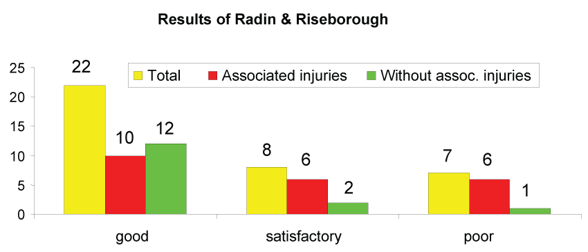
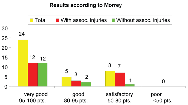
Patients showed an average Morrey score of 90 points (92; 60-100; SD 10.8). According to criteria set forth by Morrey, 24 patients (65%) received a score of very good, five patients (14%) good, and eight patients (22%) satisfactory. No patient received a poor score. According to Radin und Riseborough, 22 patients (59%) received a score of good, eight (22%) satisfactory, and the remainder (19%) poor.
In the group with associated injuries, 12 patients (55%) had very good results, three (14%) good, and seven (32%) satisfactory according to Morrey. In the group without associated injuries, 12 (80%) had very good results, two (13%) good, and one (7%) satisfactory (Figure 1, Figure 2, Figure 3 and Table 5).
.
Figure 1: Comparison of patients with and without associated injuries of the radial head after ORIF (Radin and Riseborough [24]).
View Figure 1
.
Figure 2: Comparison of patients with and without associated injuries of the radial head after ORIF [5].
View Figure 2
Rehabilitation
At the time of injury, 33 patients (89%) were employed. Three patients were homemakers and one patient was retired. Overall, 10 patients (27%) had employment that could be classified as hard labor (e.g. carpenter), 20 patients (54%) middle hard (e.g. production manager), and seven patients (19%) light (e.g. office work).

.
Figure 3: The figure shows the results of each category according to Morrey [5], The green area shows average percentual outcome.
View Figure 3
![]()
Table 5: Results of each category according to Morrey [5].
View Table 5
Length of disability
The disability interval was less than six months in 27 patients (73%), 6-12 months in 7 patients (19%), and over 12 months in three patients (8%). Twenty-eight patients (76%) continued with their work without limitations. Eight patients (22%) reported limitations. One patient (3%) who had had an unstable elbow luxation fracture was required to change jobs due to a postoperative infection which ultimately lead to an elbow arthrodesis [26].
Radiological results
A radiological examination was done in 34 patients. In five cases (15%), there was a loss of reduction. In these five patients, one patient had an instance of implant failure due to loosening of the mini-T-plate. Another patient received a reosteosynthesis with a cancellous bone graft. Consolidation took place in the remaining three who had loss of reduction and dorsal angulations up to 20°. Non-unions were diagnosed in two patients (6%). The remaining 27 patients (79%) showed adequate consolidation. Joint displacement was observed in 24 patients (71%), from which 14 (41%) had up to 1 mm, 8 patients (24%) between 1-2 mm, and two patients (6%) between 2-3 mm. Periarticular ossification was classified according to Brooker [25]. Twenty-three patients (68%) showed no periarticular ossification; five patients (15%) showed minimal, two (6%) some, und four (12%) substantial periarticular ossification. Humeroulnar osteoarthritis was diagnosed in one patient (3%), in four (12%) some, in 11 (32%) minimal, and in 18 (53%) there was no osteoarthritis in the humeroulnar joint. Ten patients (29%) showed ulnar advancement of 1.7 mm in radiographs of the wrist.
(Case 1 & Case 2)
Subjective patient evaluation
Minimal or some pain was reported by 54% of patients with 43% reporting no pain. One patient (3%) had severe pain. Thirty-eight percent of patients never had pain, 27% rarely, 19% occasionally, 14% regularly, and 3% always. Patients reported a subjective reduction in strength in 57% of cases. Nineteen patients (51%) were very content, eight (22%) content, eight (22%) satisfied, and two (5%) were not satisfied. Regarding everyday activities, 65% had no limitations, 32% had few limitations, and 3% significant limitations. A subjective reduction in wrist strength was reported in 29%. Seventy-one percent of patients reported no limitations. Twelve percent complained of pain in the wrist under strain and at rest, 14% only under substantial strain, and 74% reported no pain at all.
Complication
Osteosynthesis failure was seen in only two cases: reosteosynthesis was performed in one patient two weeks after diagnosis, and an Evolve® prosthesis was implanted in the other patient. Four patients showed periarticular ossification. One patient required an arthrolysis. Two patients experienced a loosening of a screw through the use of an external fixator. This resulted in an unstable elbow luxation fracture and an infection requiring multiple surgeries and ultimately ending in an arthrodesis. Other complications included a temporary injury to the deep branch of the radial nerve and a compression of the radial nerve requiring revisional surgery. At no point in follow-up (after 32 and 46 months) were other neurological deficits seen.
Discussion
We were able to show in our study collective that reconstruction and osteosynthesis of radial head fractures leads to overall good functional results. Twenty-nine patients (79%) were evaluated according to Morrey as good or very good, and eight patients (22%) as satisfactory. Disability was documented as less than six months in 27 patients (73%), and 6-12 months in 7 patients (19%).
Strengths of this study were the size of the patient collective and the extensive clinical examination and documentation. One possible weakness is the absence of a comparison group, which was impossible to achieve because non-dislocated fractures are conservatively treated and higher grade injuries are mostly treated with a radial head prosthetic device in our center.
In our clinic, radial head fractures are treated operatively if more than one third of the joint surface is affected and dislocation of more than two millimeters is present. AP and lateral radiographs are routinely taken as well as Greenspan images. If the fracture is multi-fragmented, an additional X-ray of the wrist is taken. A CT is performed only if a decision has to be made if operative or conservative treatment should be done (e.g. in instances of capitullum fractures).
If a fracture can be anatomically reconstructed and afterwards is functionally stable, ORIF is a possible treatment option [27]. In our centre, ORIF is always performed in combination with treating associated injuries, including the reconstruction of ligament injuries or coronoid fractures. The goal is any case to achieve a functionally stable joint with the possibility of early mobilization [27,28].
Resection of the radial head was previously believed to be the best treatment of radial head fractures [19,29,30]. Numerous studies have shown that chronic conditions can result from radial head resection, especially in patients with associated injuries, such as cubitus valgus, ulnar advancement, mobility limitations, loss of strength, and pain in the elbow [1,2,26,27,30-33]. Mutschler et al. reported chronic pain in the wrist and proximal advancement of the radius [34].
Beingesser et al. showed in a biochemical cadaver study that a radial head resection in combination with ligament injury leads to elbow instability; however, if there was no ligament injury present, resection did not apparently lead to instability [12,35].
Itamura et al. documented results for a patient collective of 24 patients with Mason II and III fractures who had no ligament injures according to clinical examination; however, according to MRT images, there was an injury to the medial ligament in 13 patients (54%), and lateral ligament in 18 patients (80%) [6]. In the case of a radial head fracture, according to our biomechanical understanding, one would expect elbow instability with chronic problems. Also it's very important to check the adjacent joint e.g. the wrist for any kind of fracture; as well for a monteggia-like-lesion or galeazzi fracture.In our collective we attained a Morrey score of very good and good for 29 Patienten (79%). Eight patients (22%) had satisfactory results. There were clearly worse results for patients according to Morrey and Radin and Riseborough if patients had associated injuries of the elbow (Figure 1 and Figure 4). The subjective evaluation of patients showed similar results: 73% had good or very good results.
.
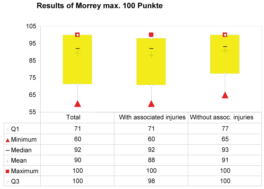
Figure 4: Comparison of patients with and without associated injuries of the radial head after ORIF (Morrey point scores [5]). 25% are less or equal to Q1; 75% are less or equal to Q3.
View Figure 4
On average, strength of the affected limb in comparison to the healthy side was reduced by 9%. Seventeen patients (46%) had a strength deficit in the injured arm compared to the healthy side and 54% of injuries affected the dominant side. A lessening of strength was reported in 57% of patients, from which there was only a small difference in strength according to our measurements with a Jamar®-Dynamometer.
Lindenhovius et al. confirmed the advantage of ORIF over radial head resection because it offered more stability and they saw a reduction in humeroulnar osteoarthritis [36]. They reported worse results for the osteosynthesis of multi-fragmented fractures and stated that a resection of the radial head was perhaps a better choice.
In a similar study, Ikeda et al. reported that patients receiving ORIF showed improved mobility and functionality of the elbow as well as more strength than in the group with a radial head resection [16].
It should, however, be emphasized that both studies were retrospective; in addition, one could assume that patients with radial head resection suffered from higher grade radial head injuries. Therefore, it is more meaningful to compare radial head resection with the use of a radial head prosthetic device. Our study was also retrospective because prospective studies, as described above, are only in certain instances possible to execute.
The percentage of radial head fractures with associated injuries is documented as being 30% in the literature [5]. We observed that the number of associated injuries substantially increased in severe radial head fractures. A fall with outstretched arm is the most common cause of injury in the literature. This does not appear to be true according to our experience when dealing with higher grade injuries. In our collective, 59% of patients had direct injures to the elbow.
Finally, in our collective there were two cases that required reosteosynthesis after loss of reduction. Both patients clearly had fractures that could be treated with ORIF.
Primary diagnostic and correct treatments are decisive for the proceeding healing process. A faulty osteosynthesis can lead to enormous periarticular ossification and even synathosis [2,34,37-40]. It should be noted that the intraoperative findings are often more complex and detailed as assumed preoperatively [6,41]. Lasting articular displacement can result in painful osteoarthritis. On the other hand, a rash resection can lead to instability of the elbow or proximal dislocation [42]. In both cases, revisional surgery is difficult: patients complain of functional limitations and persistent pain [36]. In such cases it is recommended to change to a radial head prosthetic device, or if injuries are isolated a total resection [27].
According to our results, the reconstruction of the radial head is recommended when a functionally stable result is possible. The decisive indication for ORIF is necessary for determining the proper operative methods. The restoration of a functionally stable situation is most successful in Mason II fractures and fractures with a maximum of three fragments.
Borderline indications are in complex multi-fragmented injuries in combination with mono-or polyligament damage. This is where reconstruction is limited and a prosthesis implantation is perhaps the better choice [7,43].
In our patient collective, we attempted to mobilize patients early. Our patients with no associated injuries showed good results. Reconstruction of the radial head appears to be a good method for treating simple fractures and its outcome is dependent on the presence of associated injuries.
Conclusion
Dislocated radial head fractures commonly have associated injuries. Therefore, the combination of reconstructing the radial head and repairing injured ligaments, allows for early mobilization, and is important for clinical outcome. If a functionally stable osteosynthesis cannot be performed, further possibilities include complete resection in fractures with intact ligaments, or if there is ligament damage and/or substantial instability, radial head prosthesis is recommended.
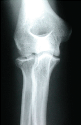
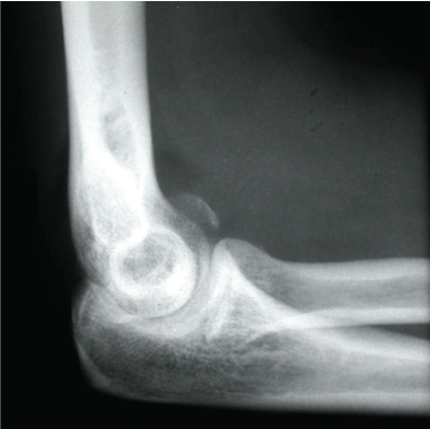
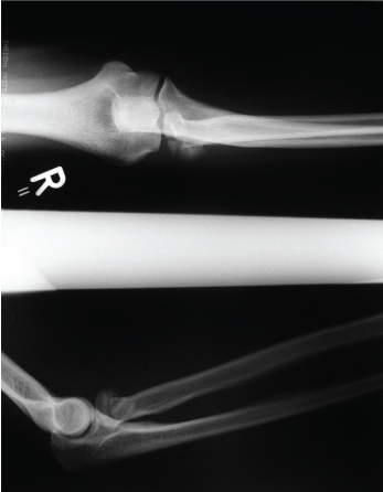
Case 1: Images 1a-d
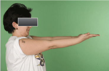
Images of a 38 year old patient after a fall while inline skating. It was a Mason II radial head fracture with a fracture of the capitullum. Follow-up after 39 months; extension/flexion in elbow 0-5-140; pronation/supination 90-0-90. The patient had a Morrey score of 93 points and good results according to R & R.
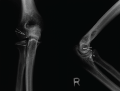
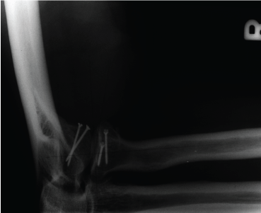
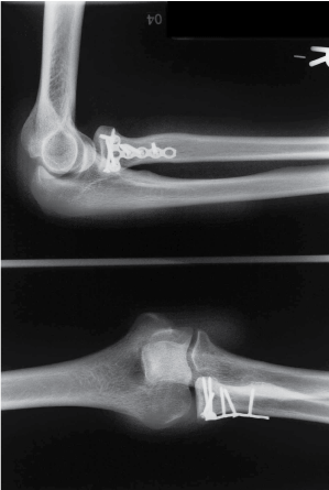
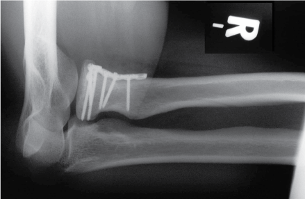
Case 2: Images 2a-g
A 34 year old patient after a fall from a chair with a Mason III fracture of the radial head and rupture of the anular ligament. Follow-up after 15 months: extension/flexion 0-0-140; pronation/supination 90-0-90. She received a score of 100 points according to Morrey and a score of good according to R & R.
Conflict of Interest
Arash Moghaddam, Tim Friedrich Raven, Peyman Kaghazian, Stefan Studier-Fischer, Tyler Swing, Paul Alfred Grützner and Bahram Biglari declare that they have no conflict of interest. Authors received no funding and have no affiliation with the company that produces materials used for osteosynthesis in this study no with a competing company. This presentation is completely product neutral.
Compliance with Ethical Requirements
The study was approved by the institutional review board "The Ethics Committee of the Landesärztekammer Rheinland-Pfalz" Number 837.322.07 (5857).
References
-
Betz A (1988) Surgical differential therapy of fracture of the radius head. Orthopade 17: 320-327.
-
Ambacher T, Maurer F, Weise K (2000) Treatment results after primary and secondary resection of the radial head]. Unfallchirurg 103: 437-443.
-
Schwarz N (1983) Conservative therapy of isolated radius head fracture--immobilization or early functional movement treatment?. Aktuelle Traumatol 13: 97-102.
-
Wallenböck E, Plecko M (1992) Complications after surgical management of radial head fractures. Unfallchirurgie 18: 339-343.
-
Morrey BF (1993) The elbow and its disorders. Saunders, Philadelphia 383-404.
-
Itamura J, Roidis N, Mirzayan R, Vaishnav S, Learch T, et al. (2005) Radial head fractures: MRI evaluation of associated injuries. J Shoulder Elbow Surg 14: 421-424.
-
Moghaddam A, Lennert A, Studier-Fischer S, Wentzensen A, Zimmermann G (2008) [Prosthesis after comminuted radial head fractures : midterm results]. Unfallchirurg 111: 997-1004.
-
Zwingmann J, Welzel M, Dovi-Akue D, Schmal H, Sudkamp NP, et al. (2013) Clinical results after different operative treatment methods of radial head and neck fractures: a systematic review and meta-analysis of clinical outcome. Injury 44: 1540-1550.
-
Koebke J (1983) A biomechanical and morphological analysis of human hand joints. Adv Anat Embryol Cell Biol 80: 1-85.
-
Putz R, Müller-Gerbl M (1988) Functional anatomy of the elbow joint. Orthopade 17: 338-346.
-
Rohlmann A, Basli A, Bergmann G (1986) Stress on the elbow joint following alloplastic joint replacement. Biomed Tech (Berl) 31: 293-302.
-
Beingessner DM, Dunning CE, Gordon KD, Johnson JA, King GJ (2005) The effect of radial head fracture size on elbow kinematics and stability. J Orthop Res 23: 210-217.
-
Charalambous CP, Stanley JK, Siddique I, Powell E, Ramamurthy C, et al. (2006) Radial head fracture in the medial collateral ligament deficient elbow; biomechanical comparison of fixation, replacement and excision in human cadavers. Injury 37: 849-853.
-
Copf F, Holz U, Schauwecker HH (1980) Biomechanical problems in elbow joint dislocations with coronoid and capitulum radii fractures (author's transl). Langenbecks Arch Chir 350: 249-254.
-
Gebauer M, Rücker AH, Barvencik F, Rueger JM (2005) Therapy for radial head fractures. Unfallchirurg 108: 657-667.
-
Ikeda M, Sugiyama K, Kang C, Takagaki T, Oka Y (2005) Comminuted fractures of the radial head. Comparison of resection and internal fixation. J Bone Joint Surg Am 87: 76-84.
-
Zimmermann G, Wagner C, Moghaddam A, Studier-Fischer S, Wentzensen A (2004) Radiusköpfchenfraktur und Ellenbogenluxation. Standards in der Unfallchirurgie. Trauma Berufskrankheit.
-
Morrey BF, Chao EY, Hui FC (1979) Biomechanical study of the elbow following excision of the radial head. J Bone Joint Surg Am 61: 63-68.
-
MASON ML (1954) Some observations on fractures of the head of the radius with a review of one hundred cases. Br J Surg 42: 123-132.
-
Greenspan A, Norman A (1982) The radial head, capitellum view: useful technique in elbow trauma. AJR Am J Roentgenol 138: 1186-1188.
-
Müller ME, Nazarian S, Koch P, Schatzker J (1990) The Comprehensive Classifikation of Fraktures of Long Bones. Berlin, Springer, Heidelberg, New York.
-
Schmickal T, Hoentzsch D, Wentzensen A (2007) A hinged external fixator for treatment of complex elbow joint injuries. Unfallchirurg 110: 320, 322-326.
-
Morrey BF (1995) Current concepts in the treatment of fractures of the radial head, the olecranon, and the coronoid. Instr Course Lect 44: 175-185.
-
Radin EL, Riseborough EJ (1966) Fractures of the radial head. A review of eighty-eight cases and analysis of the indications for excision of the radial head and non-operative treatment. J Bone Joint Surg Am 48: 1055-1064.
-
Brooker AF, Bowerman JW, Robinson RA, Riley LH Jr (1973) Ectopic ossification following total hip replacement. Incidence and a method of classification. J Bone Joint Surg Am 55: 1629-1632.
-
Moghaddam-alvandi A, Dremel E, Güven F, Heppert V, Wagner C, et al. (2010) Arthrodesis of the elbow joint. Indications, surgical technique and clinical results. Unfallchirurg 113: 300-307.
-
Ring D (2011) Radial head fracture: open reduction-internal fixation or prosthetic replacement. J Shoulder Elbow Surg 20: S107-112.
-
Kälicke T, Muhr G, Frangen TM (2007) Dislocation of the elbow with fractures of the coronoid process and radial head. Arch Orthop Trauma Surg 127: 925-931.
-
Broberg MA, Morrey BF (1986) Results of delayed excision of the radial head after fracture. J Bone Joint Surg Am 68: 669-674.
-
Keyl W (1971) Indication of radius head resection with special reference to late results of 251 fractures and luxations of the radius head. Arch Orthop Unfallchir 70: 243-260.
-
Gebauer M, Barvencik F, Rücker AH, Rueger JM (2005) Surgery for dislocated radial head fractures. Unfallchirurg 108: 669-671.
-
Morrey BF, Tanaka S, An KN (1991) Valgus stability of the elbow. A definition of primary and secondary constraints. Clin Orthop Relat Res : 187-195.
-
Walcher K (1966) Contribution to the problem of changes in the distal radio-ulnar joint following radius head resection. Arch Orthop Unfallchir 59: 316-327.
-
Mutschler W, Burri C, Rübenacker S (1990) Reconstructive surgery of malunited elbow fractures. Orthopade 19: 324-331.
-
Beingessner DM, Dunning CE, Beingessner CJ, Johnson JA, King GJ (2003) The effect of radial head fracture size on radiocapitellar joint stability. Clin Biomech (Bristol, Avon) 18: 677-681.
-
Lindenhovius AL, Felsch Q, Doornberg JN, Ring D, Kloen P (2007) Open reduction and internal fixation compared with excision for unstable displaced fractures of the radial head. J Hand Surg Am 32: 630-636.
-
Lill H, Voigt C (2004) Injuries of the elbow joint. Chirurg 75: 1037-1050.
-
Kaps HP, Niethard FU (1982) Radius head resection as a reconstructive measure of the elbow joint. Aktuelle Traumatol 12: 263-268.
-
Ikeda M, Yamashina Y, Kamimoto M, Oka Y (2003) Open reduction and internal fixation of comminuted fractures of the radial head using low-profile mini-plates. J Bone Joint Surg Br 85: 1040-1044.
-
Ikeda M, Sugiyama K, Kang C, Takagaki T, Oka Y (2006) Comminuted fractures of the radial head: comparison of resection and internal fixation. Surgical technique. J Bone Joint Surg Am 88 Suppl 1 Pt 1: 11-23.
-
Meyer-Marcotty MV, Lahoda LU, Hahn MP, Muhr G (2002) Differential therapy of radial head fracture: a critical analysis based on outcome of 53 patients. Unfallchirurg 105: 532-539.
-
Nalbantoglu U, Kocaoglu B, Gereli A, Aktas S, Guven O (2007) Open reduction and internal fixation of Mason type III radial head fractures with and without an associated elbow dislocation. J Hand Surg Am 32: 1560-1568.
-
Chen X, Wang SC, Cao LH, Yang GQ, Li M, et al. (2011) Comparison between radial head replacement and open reduction and internal fixation in clinical treatment of unstable, multi-fragmented radial head fractures. International orthopaedics 35:1071-1076.
