
Trauma Cases and Reviews
Life-Threatening Hemorrhage from Scrotal Arteriovenous Malformation Post Pelvic Fracture: A Case Report and Review of the Literature
Wei Wei*, Bryan Collier and John Ferrara
Department of Surgery, Virginia Tech Carilion School of Medicine (VTCSOM), USA
*Corresponding author: Wei Wei, MD, FACS, Department of Surgery, Carilion Clinic, VTCSOM, 2900 Lamb Circle, Suite 302, Christiansburg VA 24073, USA, E-mail: wwei@carilionclinic.org
Trauma Cases Rev, TCR-3-052, (Volume 3, Issue 1), Case Report; ISSN: 2469-5777
Received: June 13, 2016 | Accepted: March 22, 2017 | Published: March 25, 2017
Citation: Wei W, Collier B, Ferrara J (2017) Life-Threatening Hemorrhage from Scrotal Arteriovenous Malformation Post Pelvic Fracture: A Case Report and Review of the Literature. Trauma Cases Rev 3:052. 10.23937/2469-5777/1510052
Copyright: © 2017 Wei W, et al. This is an open-access article distributed under the terms of the Creative Commons Attribution License, which permits unrestricted use, distribution, and reproduction in any medium, provided the original author and source are credited.
Abstract
We report a rare case of a patient with known Child Class B type alcoholic cirrhosis who developed bleeding from an arteriovenous malformation (AVM) of scrotum after suffering a traumatic pelvic fracture. The patient subsequently developed hemorrhagic shock secondary to spontaneous bleeding from the scrotal AVM. Emergent pelvic angiogram with selective embolization was successful in controlling the bleeding. During four and half years of follow up, the patient has required two catheter directed embolization procedures for small degrees of recurrent scrotal bleeding (at four and 36 post injury months). There have been no recurrent episodes of bleeding in the sixteen months since the last embolization.
Keywords
Arteriorvenous malformation (AVM), Scrotum, Pelvic fracture
Introduction
Spontaneous hemorrhage from a scrotal arteriovenous malformation (AVM) has been scarcely reported. Herein we report the management of a trauma patient with a right pelvic fracture who developed delayed hemorrhagic shock secondary to bleeding from a scrotal AVM.
Case Report
A 41-year-old man with a known history of alcoholic cirrhosis (Child Class B type with hyperbilirubinemia, hypoalbuminemia, ascites and thrombocytopenia) was transported by ambulance to our level I trauma center emergency department as a victim of domestic violence. The patient was complaining only of severe right-sided hip pain. Vital signs and GCS were normal. The abdomen was dull to percussion and non-tender, though there was severe tenderness over the right hip. The right lower extremity was medially angulated and without neurovascular deficit. The right hemi-scrotum was mildly swollen, without tenderness or ecchymosis. The patient had neither difficulty voiding nor dysuria during the initial evaluation; urinalysis demonstrated no hematuria.
An admission CT scan noted a comminuted fracture of right acetabulum, with a retroperitoneal hematoma along the right iliopsoas muscle and paracolic gutter, extending into the right hip joint. The consensus of the trauma team was that the patient was likely developing the scrotal swelling secondary to direct trauma or fluid dissection from the right pelvic fracture, or both. The decision was made to admit the patient for close observation and in-hospital evaluation and monitoring of the hip fracture and scrotal swelling.
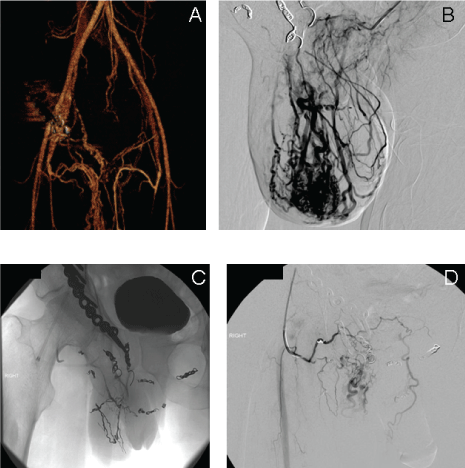
The patient underwent uneventful open reduction and internal fixation of right hip on hospital day 2. The right hemi-scrotum was noted to gradually enlarge and become ecchymotic, with the development of progressive pain during the hospital stay; local measures and mild analgesics were used to manage these symptoms. On hospital day 8, the patient was found lying in bed with his buttock area soaked in bright red blood, both confused and hypotensive (systolic blood pressure 70 mmHg). Emergent fluid and blood product resuscitation were initiated, and the patient was transferred to the intensive care unit for stabilization. Though physical examination at that time did not identify any obvious source of bleeding from the hip repair wound site, scrotum or the gastrointestinal tract, emergent esophagogastroduodenoscopy and colonoscopy were completed, with findings negative for a bleeding source. However, a pulsatile arterial bleeding site from the skin of the posterior right scrotum was later identified and controlled with skin sutures. This observation led to an emergent pelvic CT angiogram, which demonstrated a large AVM in the right hemi-scrotum, with an arterial blush. When images were compared to the admission CT scan, significantly enhanced vascularity over the right scrotum was apparent (Figure 1).
.
Figure 1: A) CT-angiogram finding from the first episode of scrotal bleeding. There was significantly increased vascularity in the right internal iliac arteries with a significant large plexus of venous circulation in the immediate vicinity of injured area. The finding suggested a significant AVM formation. Because of in lack of prior image for comparison, it was unclear if the AVM was pre-existing with acute exacerbation from pelvic fracture or caused by the right pelvic traumatic injury; B) Pelvic angiogram and embolization from the first episode of scrotal bleeding; C, D) The angiogram finding from the recurrent bleeding from the scrotal AVM.
View Figure 1
The patient then underwent selective angiogram, which confirmed the presence of a right-sided intrascrotal extratesticular AVM. A catheter-based coil embolization was performed to interrupt multiple arterial feeders from the distal branches of right internal pudendal artery and right medial femoral circumflex artery. At the termination of the procedure, persistent though minimal arterial flow to the AVM area from the distal branches of left internal pudendal artery was identified that would require serial ultrasound evaluations follow up for evidence of AVM expansion.
There was no further evidence of scrotal bleeding during the remainder of the patient's hospital stay, and clinical examination did not reveal any evidence of scrotal skin ischemia subsequent to the angiogram. The patient was thereupon transferred to an inpatient rehabilitation facility, where a two week follow up scrotal ultrasound revealed significant reduction of edema of the right scrotal wall and AVM blood supply, without testicular or epididymal abnormality.
Four months after the initial coil embolization, the patient presented to emergency department with the finding of a small amount of right side scrotal skin bleeding, which was controlled with external compression. The patient underwent an emergent pelvic angiogram, which demonstrated that the AVM had developed several collateral branches, with a dominant feeding vessel emanating from the right external pudendal artery in addition to several other small arterial feeders. The patient underwent a series of coil and gelfoam particle embolizations (Figure 1), with achievement of near-total occlusion of feeding arteries. Of note, this procedure did in fact result in a small area of scrotal skin ulceration that required wound care management. The patient was at that time considered for surgical excision of the AVM to a multidisciplinary conference that included input from specialists in the fields of urology, plastic surgery, internal medicine, and interventional radiology. The consensus was that surgical excision would be of prohibitive risk to this patient, given his severe alcoholic cirrhosis (his ascites being managed by serial paracenteses and his severe thrombocytopenia). It was elected to continue with close observation and interventional procedures as deemed necessary.
At the three year follow up evaluation, the patient was noted to have developed a small amount of bleeding from right scrotal area. Angio catheter-directed sequential alcohol and/or coil procedures were performed, and there have been no recurrent episodes of scrotal bleeding in the subsequent 16 months of follow up, although at the four year interval from the initial pelvic trauma, the patient was admitted to the hospital for symptoms of left side numbness of face, hand, and leg. The MRI and later cerebral angiogram identified a 6 mm lesion in the brainstem consistent with an intracerebellar AVM in the junction area between the pons and medulla oblangata. The neurosurgery and radiation oncology were consulted. The patient received cyber knife stereostatic radiation to brainstem AVM lesion with a plan of close follow up in the near future.
Discussion
To our best knowledge, a bleeding scrotal AVM resulting in hemorrhagic shock has never been reported in literature. A scrotal AVM is an extremely rare diagnosis in and of itself, with only several reports that document bleeding from such a lesion. In the patient reported herein, the subsequent of a brainstem AVM in addition to earlier finding of scrotal AVM suggest a hereditary pathology, possibly related to hereditary hemorrhagic telangiectasia [1] though a traumatic etiology certainly cannot be ruled out for the scrotal AVM.
The initial angiogram (Figure 1A) revealed a plethora of arterial branches and immediate filling of iliac veins. Although, lacking pre-existing imaging studies, we could not confirm if there were a preexisting scrotal AVM, we propose that trauma to a pre-existing pelvic AV fistula likely may have contributed to overt bleeding. At the very least one might speculate that the acute acetabular fracture and bony spicules would have either resulted in AV malformation/fistula as sole reason for scrotal bleed or significantly contributed to scrotal bleeding.
From review of the literature [2-5], the bleeding AVMs are either congenital or secondary to acute or remote trauma. Both catheter-based embolization and surgical excision were utilized for treatment with success (Table 1). However, the long-term success of either treatment approach is unknown, due to the rarity of the disease. Although CT angiogram is emerging as a useful tool of diagnosis of the AVM, catheter based arteriogram is considered as the gold standard for diagnosis and appropriate initial treatment. Catheter based embolization of the arterial blood supply of the bleeding scrotal AVM is usually successful in relief of symptoms, but it can at times be difficult to achieve complete obliteration of the culprit vessels. Some surgeons advocate surgical excision of the scrotal AVM as a definitive treatment [5]. However, the recurrent AVM related symptoms has been reported even at 12 years after initial successful excision [6].
![]()
Table 1: Characteristics of bleeding scrotal AVM.
View Table 1
In this current case scenario, surgical excision was proposed at the time of recurrent bleeding episode. However, due to the patient's severe comorbid condition, surgical excision was not deemed prudent. Surveillance and staged embolization in a close follow up were adopted for this patient care-with success to date.
Conclusion
Based on the review of literature in addition to our limited experience from the current case with a follow up of four and half years, we suggest that the optimal treatment of bleeding scrotal AVM should be individualized. We do believe that angiogram and catheter based embolization should be considered as the initial approach for control of the bleeding scrotal AVM. If the obliteration of blood supply of AVM is achieved, the patient can be safely followed as an outpatient with a CT or catheter based angiogram. However, surgical excision should be considered in otherwise healthy patients if embolizations fail to result in complete occlusion of the blood supply of AVM, in order to avoid high risk of recurrent bleeding.
References
-
Peacock HM, Caolo V, Jones EA (2016) Arteriovenous malformations in hereditary haemorrhagic telangiectasia: looking beyond ALK1-NOTCH interactions. Cardiovasc Res 109: 196-203.
-
Jaganathan S, Gamanagatti S, Mukund A, Dhar A (2011) Bleeding scrotal vascular lesions: interventional management with transcatheter embolization. Cardiovasc Intervent Radiol 2: S113-S116.
-
Hatten BW, Bryant E (2012) Bleeding scrotal arteriovenous malformation. J Emerg Med 42: e133-e135.
-
Kang TW, Choi YD, Jeong YY, Kwon DD, Park K, et al. (2004) Intrascrotal extratesticular arteriovenous malformation. Urology 64: 590.
-
Sule JD, Lemmers MJ, Barry JM (1993) Scrotal arteriovenous malformation: case report and literature review. J Uro 150: 1917-1919.
-
Bandi G, Bianco F, Dhabuwala CB (2004) Recurrent scrotal arteriovenous malformation. J Urol 171: 1628.
