Granular cell tumors are unusual, typically benign tumors deriving from Schwann cells. They can arise of tissues anywhere in the body and can rarely involve the perianal tissues. We discuss the case of a granular cell tumor involving the external anal sphincter.
This is a case report with review of the literature.
A 52-year-old woman presented with a growing perianal mass found on exam and imaging to involve the anal sphincter causing impaired continence of stool. The mass was biopsy proven to be a granular cell tumor. Local resection with overlapping sphincteroplasty repair was performed. Continence was preserved. We review histopathologic and imaging features of granular cell tumors as well as the natural history of these lesions.
Granular cell tumors can very rarely involve the anal sphincters. Adequate resection is important for local control. This can be achieved with preservation of continence.
Perianal, Granular cell tumor, External anal sphincter, Anorectal, Schwann cell tumor
Granular cell tumors are rare, generally benign, neoplasms derived from Schwann cells that can arise anywhere in the body. The perianal region is a rare location for granular cell tumors, and it is even more unusual to present with involvement of the anal sphincter muscles. We present a case of a granular cell tumor involving the external anal sphincter. We discuss management of this unusual neoplasm as well as review the pertinent literature.
The patient is a 52-year-old female who presented for evaluation of a newly identified mass near her anus. She was undergoing evaluation of recently diagnosed multiple sclerosis, and during physical examination was noted to have a lesion at the anal verge. She reported that she may have been aware of a mass near her anus for the last year. It had been slowly growing over time from about one centimeter to about four centimeters. She denied pain associated with the mass. It did not initially compromise her continence, but as the mass grew, she had begun to develop seepage and occasional incontinence of solid stool. Her obstetric history was significant for a single pregnancy and vaginal delivery with an episiotomy. Her medical and surgical history was remarkable only for multiple sclerosis and Class III obesity with BMI 45 kg/(m2). There was no significant family history.
On exam, she was noted to have a very firm, partially mobile lesion palpable at the anal verge in the right anterior position. This lesion was nodular and discrete but was not exophytic. The skin overlying and surrounding the lesion was hyperpigmented and intact except for a small cleft in the lesion. On palpation, the lesion felt as though it was affixed to and possibly arising from the external anal sphincter.
A punch biopsy was taken of the lesion. Overlying squamous mucosa was normal. The underlying tumor revealed large cells with abundant fine granular cytoplasm. There were small, centrally-located nuclei and no mitotic figures or atypia noted on hematoxylin-eosin staining. Immunostaining for the S100 protein highlighted the nuclei of the tumor cells with only weak staining of the cytoplasm, which is consistent with a granular cell tumor (Figure 1).
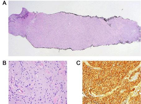 Figure 1: Micrographs of punch biopsy of perianal granular cell tumor.
A) Low magnification view. Normal overlying perianal squamous mucosa is seen to the left of the section; B) High magnification (20X) H&E stain of granular cell tumor. There are abundant large cells with abundant fine granular cytoplasm and small, centrally located nuclei. No mitotic figures or cellular atypia are apparent; C) High magnification view (20X) of immunostaining of tumor cells for S100 protein which highlights the nuclei while only weakly staining the cytoplasm's. View Figure 1
She underwent pelvic magnetic resonance imaging, which showed a heterogeneously enhancing soft tissue perianal mass with restricted diffusion. The lesion did appear to involve both the internal and external anal sphincters with invasion into the ischioanal fat. There were also mildly enlarged pelvic lymph nodes (Figure 2A).
On endoanal ultrasound, the mass appeared relatively hypoechoic with indistinct borders, with clear involvement of the external anal sphincter. Flexible sigmoidoscopy and endoscopic ultrasound were also performed to further evaluate the pelvic lymph nodes; cytology on fine needle aspiration was non-diagnostic (Figure 2B).
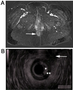 Figure 2: Imaging of perianal granular cell tumor involving external anal sphincter.
A) Magnetic resonance imaging of perianal granular cell tumor (white arrow). The tumor is a heterogeneously enhancing soft tissue mass with restricted diffusion which involves the sphincter muscles and invades ischioanal fat; B) Endoanal ultrasound of perianal granular cell tumor (white arrow). The mass is hypoechoic relative to surrounding ischioanal fat with indistinct borders. It involves the external anal sphincter (*indicates internal anal sphincter; **indicates external anal sphincter). View Figure 2
Management options were discussed extensively with the patient. Despite the relatively indolent nature of granular cell tumors, the tumor was already involving the sphincter and compromising her continence. She was counseled that a watch-and-wait approach would likely result in progression of incontinence and anal pain. Abdominoperineal resection was also discussed as definitive therapy due to sphincter involvement. The patient was an otherwise healthy 52-year-old woman, and was not interested in a permanent colostomy if there were other reasonable courses of action. Ultimately, she was offered local excision with overlapping sphincteroplasty and careful surveillance, and this was the patient's preferred approach.
The patient was taken to the operating room and placed in the prone position after induction of general anesthesia. A circumanal incision marked to include the lesion with a gross margin was carried through the subcutaneous tissue and into the ischiorectal fat (Figure 3A). A firm, well-circumscribed mass which involved 30% of the external anal sphincter up to a depth of 2.5 cm was identified (Figure 3B). Intraoperatively the internal anal sphincter was dissected free and preserved intact (Figure 3C). The healthy external anal sphincter adjacent to the tumor was mobilized both medially and laterally (Figure 3D). The external sphincter was then divided laterally and posteriorly to allow for a margin on the tumor. The ends were tagged to facilitate repair (Figure 3E). A deep margin was obtained on the external sphincter and the lesion was then excised. The margins were grossly evident, and the deepest portion of the external anal sphincter was spared. An overlapping sphincteroplasty of the external anal sphincter was then performed using 2-0 absorbable polyglactin suture (Figure 3F). The soft tissue defect was closed in layers, and a penrose drain was left to help evacuate the resection bed (Figure 4A). Postoperatively the patient had wound breakdown of the soft tissue closure that healed with local wound care (Figure 4B, Figure 4C and Figure 4D). The sphincteroplasty remained intact. The patient initially had urgency with frequent fecal incontinence because of inability to defer defecation, but this improved as the wound healed. By eight months postoperatively, she reported one or two episodes of fecal incontinence per month due to urgency.
 Figure 3: Resection of perianal granular cell tumor involving external anal sphincter. The patient is in prone position.,
A) Retraction sutures are placed, and the circumanal incision including skin overlying tumor which is in the right anterior position is marked; B) The incision is carried through the subcutaneous tissue and into the ischiorectal fat so that the firm, well-circumscribed mass was completely dissected free; C) The internal anal sphincter is dissected free and intact. This is being retracted posteriorly and toward the left in this panel, while the mass and involved external anal sphincter is being retracted anteriorly and to the right; D) The uninvolved external sphincter was mobilized medially and laterally prior to dividing the muscle to remove the tumor en bloc with an adequate margin of grossly uninvolved tissue; E) The end of the external anal sphincter with tagged with suture; F) The appearance of the completed overlapping sphincteroplasty. View Figure 3
 Figure 4: Wound healing following resection of perianal granular cell tumor involving the external anal sphincter with overlapping sphincteroplasty.
A) Wound at the completion of case. Soft tissue defect was closed in layers and Penrose drain was left to ensure ongoing drainage; B) By postoperative day 8, the drain has been removed and the wound overlying the soft tissue closure has dehisced. The sphincteroplasty is still intact. Local wound care and dressing changes are initiated; C) Postoperative day 45. The wound has decreased in size and depth; D) Postoperative day 80. The wound is almost entirely healed except a small area of granulation tissue anteriorly. View Figure 4
The final pathology report revealed a 4.7 cm granular cell tumor. The deep resection margin was focally involved by tumor cells. There was no evidence of gross tumor recurrence at 3 months postoperatively. Repeat MRI at eight months postoperatively revealed a 1.2 × 1 cm soft tissue mass in the right ischioanal fossa resection bed concerning for residual or recurrent tumor. The mildly enlarged lymph nodes that had been noted on the original MRI were stable. On physical exam, no residual nodule could be palpated corresponding to the soft tissue mass seen on MRI. A repeat MRI will be obtained at 12 months, and if this soft tissue lesion continues to enlarge, a limited transanal resection will be performed.
Granular cell tumors (GCTs) are rare tumors that can occur in any location. They were first described in 1926 by Abrikossoff and were initially thought to arise from degenerating striated muscle [1]. Since that time, the consensus is that these tumors are derived from Schwann cells [2,3].
GTCs have been described in a multitude of locations throughout the body. They commonly arise in the oral cavity, with the tongue being the most common location; skin, particularly in the subcutaneous tissues of the chest and upper extremities; and the gastrointestinal tract from the esophagus to the anus and perianal tissues. Additionally, GCTs have been described in the orbit; mastoid region; nervous system; respiratory tract; breast; biliary system; and genital tract [1,3-5]. A case series of 50 GCTs from any location within the body was collected from a cancer center. In this cohort, the median age was 47, and 62% were female. The majority of patients were white (64%), and 94% had unifocal tumors. Most tumors (86%) were benign by histopathological and clinical criteria [5]. In contrast, a case series examining the distribution of GCTs in the gastrointestinal tract found that of the 74 tumors, 24 arose from the esophagus, 8 from the stomach, 3 from small bowel, 4 from the appendix, 20 from the colon and rectum, and 16 from the anus or perianal tissues [4]. Gastrointestinal GCTs were most commonly diagnosed between the third and fifth decades of life, and there was a slight female preponderance. None of the gastrointestinal GCTs in either of these series exhibited malignant behavior [4-6].
GCTs in the perianal area typically present as an incidentally found nodules or polypoid lesions. If patients have noticed them, they report a slowly growing firm nodule near the anus. Some patients report pain, but many nodules are painless. They can also be found incidentally during evaluation and treatment of hemorrhoids or hematochezia. On exam, lesions are described as firm and nodular. Often, they are deep to the dermis and freely mobile, but can be affixed to the overlying skin [1,3-13].
Imaging features of granular cell tumors tend to be nonspecific. CT scans show well-circumscribed soft-tissue masses that do not have calcifications. Ultrasound imaging of biliary GCTs reveals heterogeneous, mildly hyperechoic lesions that may have faint posterior shadowing [14]. Our endoanal ultrasound images showed the tumor to be heterogeneous and hypoechoic relative to surrounding ischioanal fat. On MRI, GCTs are heterogeneous with low signal intensity on T1 weighted images and high signal intensity on T2 weighted images [6].
GCTs exhibit a characteristic appearance on histopathologic examination. They typically have plump ovoid to polygonal cells with coarse eosinophilic granules in the cytoplasm. The nuclei are small and round to ovoid with distinct nucleoli. Mitotic figures and pleomorphism are usually absent [3,15]. The overlying epidermis can exhibit pseudoepitheliomatous hyperplasia. Immunohistochemical staining is key for diagnosis. GCTs stain strongly positive for S100 protein, neuron-specific enolase, and vimentin. They stain negative for markers specific for epithelial, melanocytic, muscle, endothelial, and glial cells, all of which support their Schwann cell origin [3,15].
Treatment of perianal GCTs consists of complete local excision. The majority of tumors are benign, and almost every reported tumor has not recurred after adequate local excision. However, local recurrence is possible [9], and GCTs can exhibit malignant behavior [12]. While not specific to perianal tumors, features suspicious for malignant behavior include cell necrosis, spindling, pleomorphism, increased mitotic activity (> 2 mitoses/10 HPF at 200x magnification, vesicular nuclei with large nucleoli, and high nuclear to cytoplasmic ratio [16]. The larger number of these criteria that are met, the higher the malignant potential of the tumor. Patients should be carefully monitored after excision for recurrence and for lymphatic or hematogenous spread. While it is very rare for perianal GCTs to exhibit malignant behavior, when metastases occur, there are no reliably effective salvage strategies described [12,17].
Delineating the relationship between GCTs and surrounding tissue is of paramount importance in surgical planning. While most perianal GCTs are in the subcutaneous tissues in or around the anal canal, we report here the fourth case of a GCT involving the anal sphincter complex in the literature [7-9]. Complete local excision is key to management of GCTs, but preservation of continence must be taken into consideration when the sphincter complex is involved. The specific demographics, excision and reconstruction strategies, and outcomes of GCTs involving the anal sphincters are summarized in Table 1. Suspicion that the anal sphincter complex is involved by perianal GCT should prompt careful imaging workup in order to inform preoperative discussions with patients about risks and outcomes of surgery.
Table 1: Summary of cases of granular cell tumors involving the anal sphincters: Demographics, treatments, and outcomes. View Table 1
Perianal GCT is a rare entity that is typically benign and can be adequately treated with local excision and careful follow-up to monitor for local recurrences. Malignant behavior from perianal GCTs is exceedingly rare. However, GCTs can involve the anal sphincter complex. In these instances, careful preoperative workup should be undertaken to ascertain the extent of sphincter involvement so that the patient can be informed about the likelihood that an adequate resection can be performed while still preserving continence. Fortunately, sphincter involvement by GCTs is rare, and the few cases that have been reported, including this case, describe excellent functional outcomes.
Thank you to Dr. Shu-Yuan Xiao with the University of Chicago Department of Pathology for assistance with histopathology. Thank you to Dr. Brian Olson for assistance with production of figures.