Deep brain stimulation (DBS) is an established treatment for advanced Parkinson’s disease, and although generally safe, complications related to the implantable impulse generators (IPGs), such as infection, skin erosion, or device malfunction, can arise. Malignancy arising at the IPG implantation site is exceedingly rare, with few cases previously reported, related to pacemakers IPGs. We present case of invasive ductal breast carcinoma developing adjacent to a DBS IPG. A 77-year-old woman with Parkinson’s disease underwent bilateral subthalamic nucleus DBS implantation in 2013. In 2020, she was diagnosed with stage 3 ER+/HER2- ductal invasive carcinoma in the left breast. She underwent radical mastectomy and was placed on antioestrogen therapy. Two years later, erythema and edema over the IPG site led to a presumptive diagnosis of infection, treated unsuccessfully with antibiotics and device relocation. Further evaluation revealed cutaneous metastatic recurrence localized to the IPG region, without systemic spread. The disease progressed locally, and the patient died a year later. This case underscores a rare but critical diagnostic challenge-malignancy mimicking IPG pocket infection. Clinicians should maintain a high index of suspicion for malignancy in atypical IPG site presentations, especially in patients with a prior cancer history.
Deep brain stimulation, Metastasis, Malignancy, Ductal breast cancer, Implantable pulse generator
CT: Computer Tomography; DBS: Deep Brain Stimulation; IPG: Implantable Impulse Generators; STN: Subthalamic Nucleus
Deep brain stimulation (DBS) is a neurosurgical treatment, approved for several neurological and psychiatric conditions that are in an advanced stage, or refractory to oral medications. It is approved for the management of advanced Parkinson’s disease, with well-established improvement of tremor, rigidity, motor fluctuations and other and non-motor complications of this disease [1]. Deep brain stimulation procedures have consistently increased over the years [2] and, despite their miniaturization and sophistication, DBS systems share a common set of components, powered by an implantable pulse generator (IPG) [2]. These devices, after implantation, can be related to several early or delayed complications related to infection, skin erosion, flipping (Twiddler’s syndrome), wound dehiscence, malfunction [2], which can cause significant morbidity and/or mortality. Rare cases of malignant neoplasms around implantable pulse generators adjacent to cardiac pacemakers have been described. We report here a case of an invasive ductal breast carcinoma arising in the subcutaneous tissue adjacent to the DBS’s IPG location.
A 77-year-old woman with a known history of Parkinson’s disease, first diagnosed at 47 years of age, suffering from both motor and non-motor disease complications, was submitted to bilateral subthalamic nucleus (STN) deep brain stimulation (DBS) surgery in 2013, with improvement of motor symptoms. She was also implanted with a subcutaneous left thoracic chest pulse generator, whose battery was replaced in 2018 with a minor surgical procedure, with no related complications.
In 2020, she developed a petrous lump with 2cm of diameter, on the left superior quadrant of the left breast, which, after thorough evaluation and staging, was classified as a ductal invasive breast carcinoma that had already metastasized to her left axillary lymphatic ganglia (Stage 3, grade 2, pT2 pN2a) expressing hormone receptors (ER+) but not HER+ receptors, with a Ki67 of 40%. Following this diagnosis, she was submitted to a radical left mastectomy in 2020, proposed for left breast radiotherapy, which she didn’t start, and was started under prolonged systemic antioestrogen therapy, with letrozole. There was no evidence of metastatic spreading lesions on bone scintigraphy and also CT scan of thorax, abdomen and pelvis. There was no evidence of progression in the following months of follow-up. Two years after surgery completion she developed left thoracic skin rubor and edema, just above the device’s implantation cavity site, with no signs of fluctuation (Figure 1). Initially, a subcutaneous tissue infection was assumed, and she was started on antibiotics (8-day course of intravenous flucloxacillin). There was no improvement of symptoms, so the surgical site was opened and cleaned, and the pulse generator implanted on the right upper thoracic subcutaneous tissue. The disease continued to progress locally, on the left and on the right side, and a subcutaneous metastatic disease progression of her original invasive breast cancer was diagnosed, with no signs of remote disease progression. The patient’s condition worsened during the following year, she was referenced to palliative care and eventually died due to the progression of her illness.
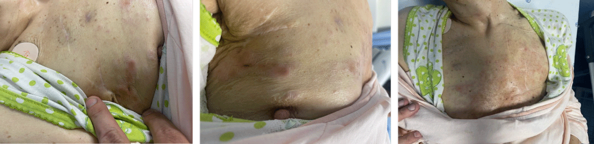 Figure 1: Pictures showing breast carcinoma skin metastasis over chest implantable device site.
View Figure 1
Figure 1: Pictures showing breast carcinoma skin metastasis over chest implantable device site.
View Figure 1
Malignancies arising from the implantable pulse generator pocket site are a very infrequent local complication, being previously described as related to pacemakers IPGs [3]. Additionally, reports of tumours arising primarily in the pocket of these devices are even rarer in the medical literature, according to Morais, et al.
Like most of the patients described before, in this case, malignancy presentation was with a petrous lump over the location of the IPG, associated with local inflammatory signs that appeared seven years after the device’s first implantation and after two years of its battery revision. The disease progressed locally, with the development of cutaneous and subcutaneous nodules, with no associated local or remote lymphadenopathy.
The process of formation of malignant lesions around IPGs is still not precisely understood. One hypothesis might be related to the release of metal ions by the generator which can cause toxicity and pro-inflammatory and oxidative stress effects mediated by several interleukins and chemokines (TNF-α, TGF-β, etc.) that might also relate to genomic instability or cancer related genetic mutations in the tissues around the IPG [3]. Another hypothesis might be related to the chronic inflammation provoked by IPGs chronic mechanical irritation or by its electrical stimulation, which might cause chronic immune cell activation, cellular damage and promote cancer cell migration due to electrical activity (galvanotaxis) [4,5].
Although rare, several breast malignancies have been previously reported as an IPG invasive tumour [6-11]. Different and heterogenous management strategies were used following the malignancy diagnosis, some including surgery, chemotherapy and radiotherapy, according to the different histopathology,
In our case, since this diagnosis was not initially suspected and since the patient had her oncological disease controlled after surgery and antioestrogen therapy, it was assumed as a local infection complication, so she was just submitted to a cycle of intravenous antibiotics and surgical cleaning of the IPG site. There was no specialized local tumour removal during this procedure, and the device was implanted on the contralateral side. She was kept under antioestrogen therapy, but no cycle of chemotherapy or radiotherapy was done which, also might relate to the subsequent local progression of the disease. Due to the rarity of these tumours, there is a lack of consensus on the approach to their correct diagnosis and subsequent treatment. This case also highlights that malignancies arising from IPG implantation sites might be mistaken for pocket site infections, and a lower suspicion threshold for this occurrence is needed to correctly and promptly identify this severe disease. Given the rarity of such presentations, diagnosis is often delayed, and standardized management strategies are lacking. Further research is needed to elucidate pathophysiological mechanisms and guide clinical decision-making.
This work has not received any contribution, grant or scholarship.
The authors declare that they have followed the protocols of their work center on the publication of data from patients.
The authors declare that they have contributed in similar ways in the design and writing of this work.