Donor surplus human oocytes were transferred to adhesive slides. Oocytes were fixed with 1% paraformaldehyde in PBS. For permeabilization, cells were incubated in 0.1% Triton X-100 (Sigma) plus 0.1% sodium citrate in PBS. Cells were then incubated with 5% Bovine Serum Albumin (BSA; Sigma) to inhibit non-specific binding. Afterwards cells were incubated with a primary monoclonal antibody against human ZP1 (ZP1: sc-365435; Santa Cruz Biotechnology, Santa Cruz, California) for 1 h (37 ºC). For each experiment, a negative control was included. A secondary antibody anti-mouse FITC (sc-516140) conjugated with fluorescein isothiocyanate (FITC; Santa Cruz Biotechnology) was applied to cells for 45 min. Oocytes were not counterstained with DAPI and observed in PBS. Cells were observed in an epifluorescence microscope (Eclipse E400; Nikon, Tokyo, Japan). Experiences were performed in triplicate (Figure 1).
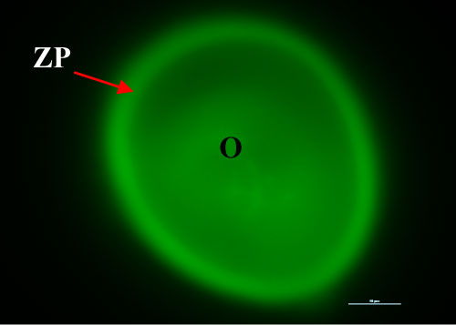
Figure 1: ZP: zona pellucida; O: oocyte. Bar: 10 µm.