Dysuria, Lithiasis, Ultrasound
A 65-year-old male, hypertensive and non-insulin-dependent type 2 diabetes mellitus as the only personal history of interest, who came to the Emergency Department of our hospital with micturition symptoms consisting of dysuria and pollakiuria associated with mild hematuria and hypogastric pain of 14 days of evolution. The patient did not report fever or any other associated symptoms. In addition, he told us that, at first, he had gone to his Health Center and consulted his Primary Care Physician for the same reason, who had prescribed antibiotic treatment because of a suspected urinary tract infection with poor therapeutic response.
During the physical examination, we found a patient with a good general appearance, hemodynamically stable although presenting arterial hypertension of 174/96 mmHg, not tachycardic (HR 80 bpm), afebrile and with saturation within the normal range (temperature 36.6 ℃, SO2 97% basal). Cardiac and pulmonary auscultation showed no pathological findings. As for the abdominal examination, we found preserved hydroaerial sounds, the abdomen was soft, depressible, not painful to deep palpation, no masses or megaliths, no signs of peritoneal irritation.
One of the first complementary tests requested after the examination of our patient is an abdominal X-ray where two superimposed radiopaque images difficult to determine were observed, so, while waiting for the rest of the results, we took the initiative to perform an abdominal ultrasound on the patient where we observed two hyperechogenic images of 6 and 4.5 cm in length, respectively, with posterior acoustic shadow compatible with bladder lithiasis (Figure 1).
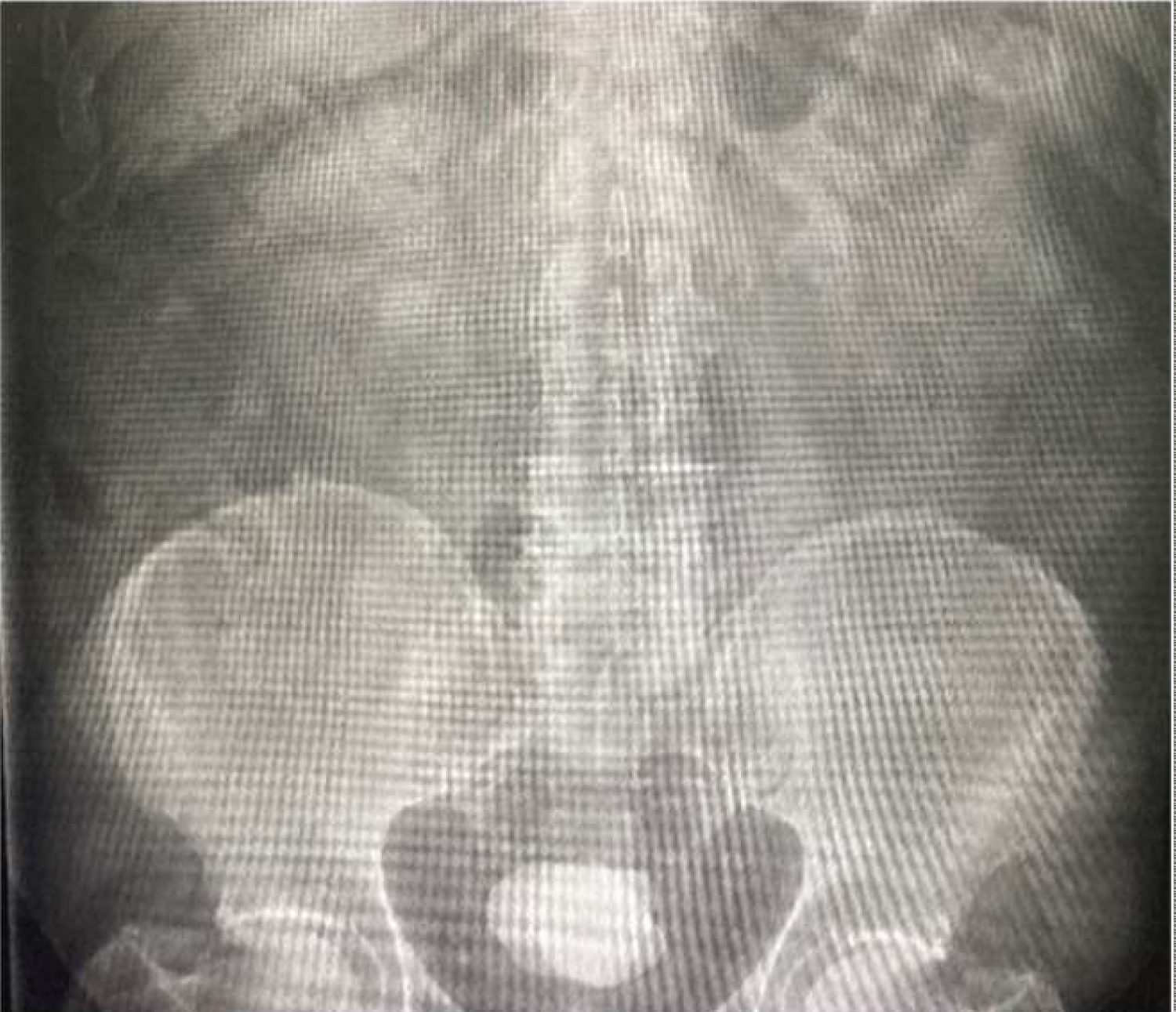 Figure 1: We took the initiative to perform an abdominal ultrasound on the patient where we observed two hyperechogenic images of 6 and 4.5 cm in length, respectively, with posterior acoustic shadow compatible with bladder lithiasis.
View Figure 1
Figure 1: We took the initiative to perform an abdominal ultrasound on the patient where we observed two hyperechogenic images of 6 and 4.5 cm in length, respectively, with posterior acoustic shadow compatible with bladder lithiasis.
View Figure 1
After these findings, it was decided to consult with the Urology Department, who confirmed the diagnosis and, in a short time, the patient underwent surgery with extraction of both bladder stones, resolving the condition for which he had consulted our Emergency Department (Figure 2).
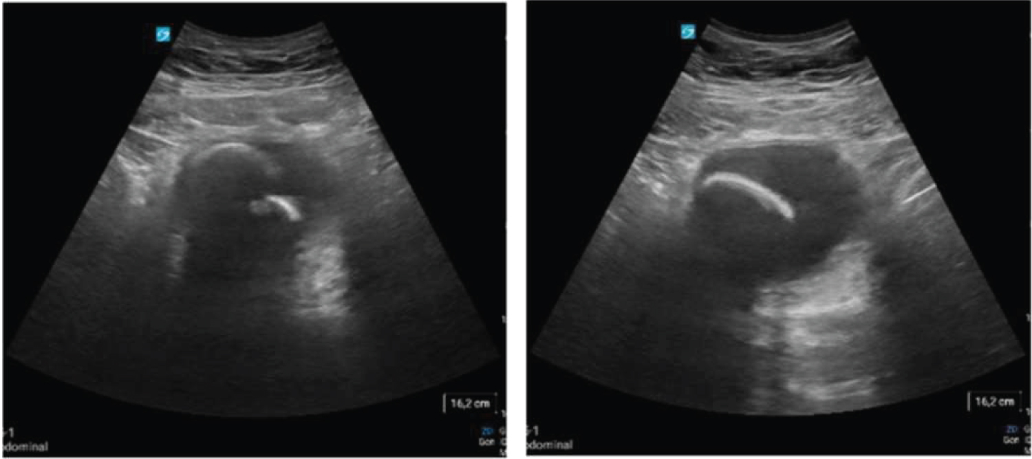 Figure 2: After these findings, it was decided to consult with the Urology Department, who confirmed the diagnosis and, in a short time, the patient underwent surgery with extraction of both bladder stones, resolving the condition for which he had consulted our Emergency Department.
View Figure 2
Figure 2: After these findings, it was decided to consult with the Urology Department, who confirmed the diagnosis and, in a short time, the patient underwent surgery with extraction of both bladder stones, resolving the condition for which he had consulted our Emergency Department.
View Figure 2
The Emergency Department receives a large number of patients every day and many of them come repeatedly after a first visit in which the problem for which they came in the first instance was not solved. It is important to reformulate the questions already asked and to evaluate other diagnostic options for repeated consultations or initial therapeutic failures, since we could be repeatedly erring in the initial clinical judgment, subjecting the patient to inadequate treatment with the consequent risk of adverse effects and the economic expense that this entails.
Fortunately, in this Service we have access to different complementary tests with relatively fast results for this purpose and we will have to evaluate which of them to perform depending on our diagnostic suspicion.
One of them is ultrasound, a tool available today in all emergency departments nationwide. This test is very useful for clarifying the possible diagnosis of a large number of pathologies for which the patient may be consulted. In addition to being useful, it is a harmless and low-cost technique with a theoretically easy and fast learning curve that will allow us to speed up the diagnostic and therapeutic process of the possible pathologies for which we may be consulted (Figure 3) [1,2].
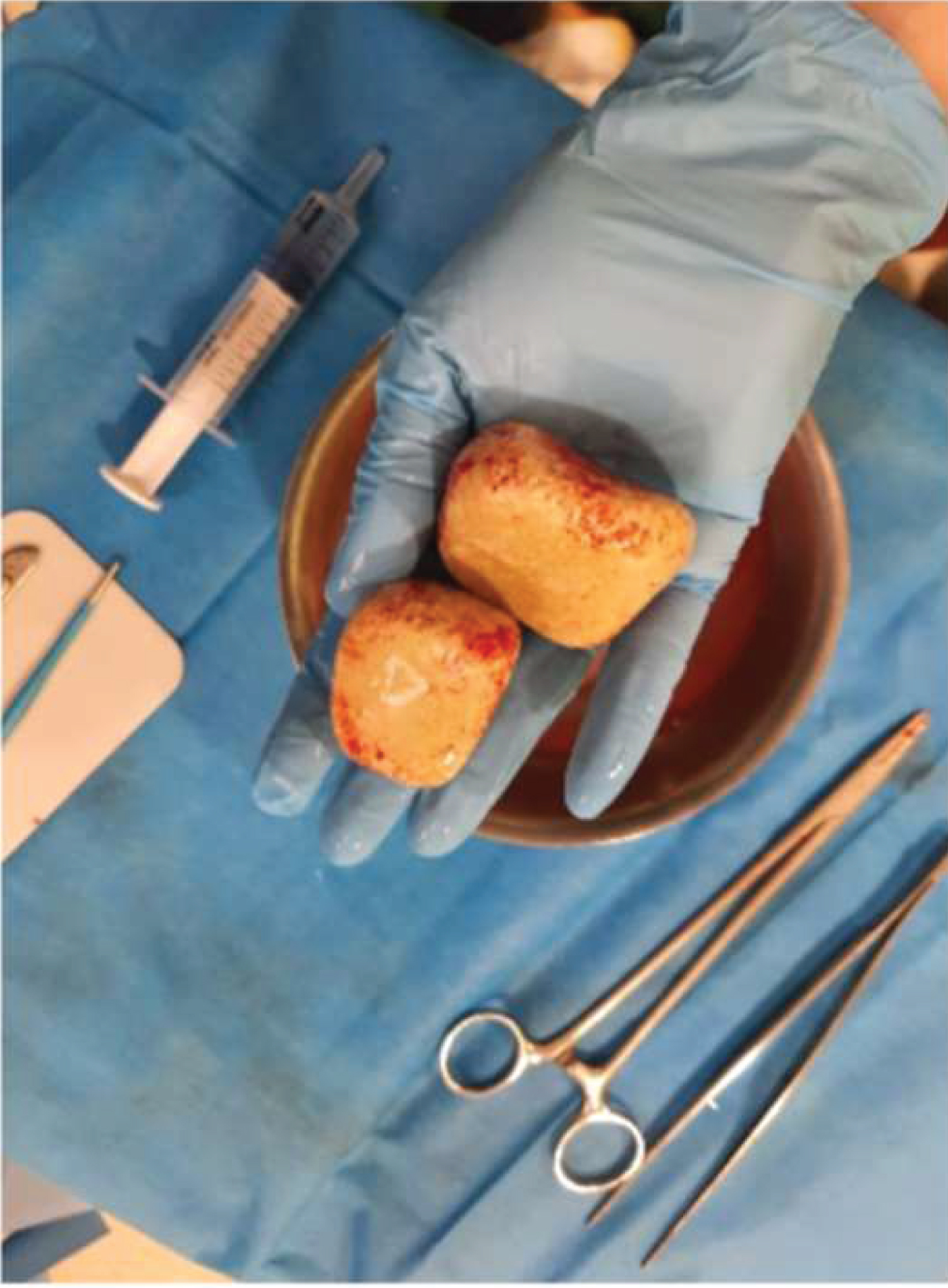 Figure 3: In addition to being useful, it is a harmless and low-cost technique with a theoretically easy and fast learning curve that will allow us to speed up the diagnostic and therapeutic process of the possible pathologies for which we may be consulted.
View Figure 3
Figure 3: In addition to being useful, it is a harmless and low-cost technique with a theoretically easy and fast learning curve that will allow us to speed up the diagnostic and therapeutic process of the possible pathologies for which we may be consulted.
View Figure 3