Background: Modular intercalary endoprostheses is a potential reconstructive option infrequently studied for diaphyseal defects of long bone. The purpose of this study was to examine the 1) Method of failure rate of revision after reconstruction with modular intercalary endoprostheses based on the anatomic site and 2) Describe the functional status of the patient and use of assistive devices with ambulation.
Methods: A retrospective chart review was performed on patients with modular intercalary endoprosthesis from 2005-2019. Inclusion criteria included long bone defects secondary to tumor resection, trauma, or infection, and treated with intercalary or knee spanning endoprosthesis in the primary or revision setting. Ambulatory status, complications, and reoperations were collected and analyzed using descriptive statistics.
Results: Nine patients out of twelve were included with three femur prostheses, three tibia prostheses, and three knee spanning arthrodesis. Mean age was 46-years-old and mean follow-up was 52 months. The four complications included structural failure and aseptic loosening in a femoral prosthesis, soft tissue failure (Type I) in a tibia prosthesis, and a local wound infection of one knee arthrodesis. All seven patients with reported ambulatory status were ambulatory at final follow-up.
Conclusion: Our study demonstrates that modular intercalary endoprosthesis is a reconstruction option that can be used for defects after tumor resection, trauma, or infection. These data warrant further investigation into the use of an intercalary endoprosthesis for patients with diaphyseal defects of the long bone.
Joint replacement, Tumour, Trauma, General
Treatment of diaphyseal defects due to tumor resection, trauma or infections is a challenging reconstructive problem. The goals of reconstruction includes limb salvage, restoration of limb length and alignment, preservation of adjacent joints if possible, early weight-bearing, and implant durability [1]. Various reconstruction techniques have been described for segmental diaphyseal defects of long bones, including biologic reconstruction with vascularized fibular autografts [2,3] intercalary allografts, [4-6] a combination of vascularized graft and allograft [7,8], distraction osteogenesis, [9] and modular intercalary endoprosthesis [1].
Vascularized fibular autografts can be used in smaller reconstructions; however, they require an initial prolonged period of non-weight bearing, are high risk for fracture, and are associated with donor-site morbidity [10,11]. Allografts have been a popular reconstructive option, offering the potential of biologic reconstruction without donor site morbidity, with a survival rate of 76-84% at long-term follow up [4-6,12]. However, allograft reconstructions are predisposed to delayed union or nonunion, infection, especially in patients who require chemotherapy or radiation, and fracture, which can be a challenging complication to treat with low rates of subsequent healing [4-6,8,12].
Intercalary endoprostheses are a reconstructive option that allows for early weight-bearing and implant fixation that is not affected by chemotherapy and radiation. Complications most commonly include aseptic loosening, mechanical failure, and infection, similar to other endoprosthetic reconstructions. However, there are limited number of studies that report outcomes after reconstruction with an intercalary endoprosthesis [1,11,13-19].
The purpose of this study was to 1) Understand the complication profile and failure mechanisms of patients undergoing intercalary endoprosthesis reconstruction; 2) Determine if complications or failures differed by anatomic site; 3) Define the rate of revision surgeries in patients undergoing intercalary endoprosthesis reconstruction; and 4) Describe the ambulatory status of these patients postoperatively, and whether they require assistive devices.
Following IRB approval, we retrospectively reviewed the records of patients who underwent endoprosthesis reconstruction by a fellowship trained musculoskeletal oncologist from April 2005 to July 2019. The indications for the use of intercalary endoprosthesis included tumor resection, trauma, or infection in the primary or revision setting in all lower extremity long bones (Figure 1). Intercalary reconstructions spanning the knee joint were included in this study. Exclusion criteria were patients without operative reports.
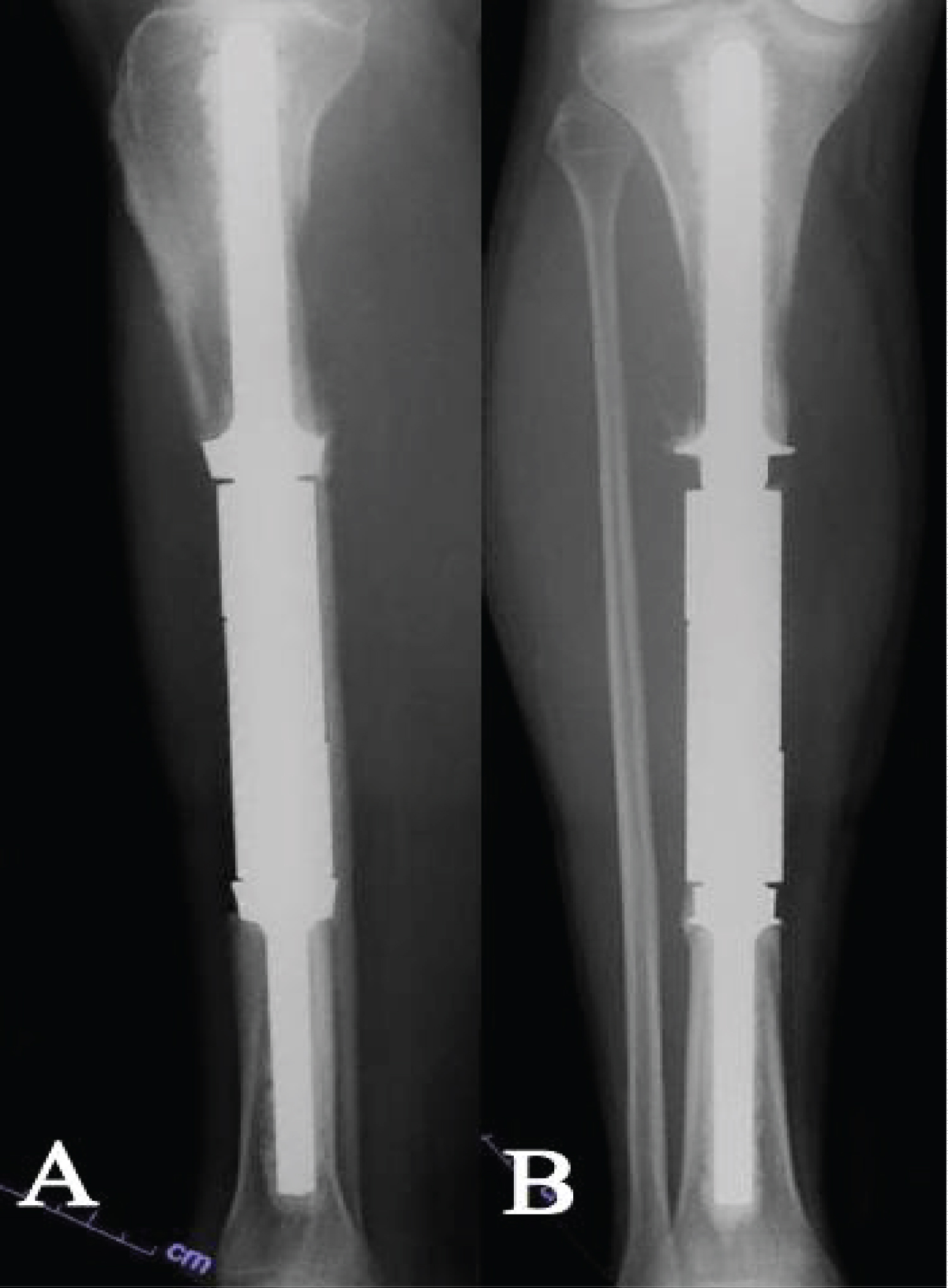 Figure 1: Anteroposterior (A) and lateral (B) radiographs of case 4 post-operatively.
View Figure 1
Figure 1: Anteroposterior (A) and lateral (B) radiographs of case 4 post-operatively.
View Figure 1
We recorded patient demographics, indication for surgery, malignancy stage if applicable, complications and method of failure according to the Henderson classification: Type I (soft tissue failure), Type II (aseptic loosening), Type III (structural), Type IV (infection), and Type V (tumor progression) [20]. The length of resection, stem lengths and intercalary body length were recorded. In patients undergoing revision surgery, the length of the resection was defined as the length of the implant or bone graft removed.
We identified 12 patients who underwent limb salvage with intercalary endoprostheses. Three patients were excluded due to missing operative reports. An example of a tibial intercalary endoprosthetic reconstruction is shown in Figure 1. Patient demographics and implant data are listed in Table 1. The mean age was 46 years (29 to 76); six patients (66.6%) were male, and the average BMI was 28.7 kg/m 2 . The intercalary endoprostheses included three femoral prostheses, three tibial prostheses, and three knee spanning arthrodeses. The mean follow up was 52 months (0 to 188).
Table 1: Details of patient and tumor characteristics, length of resection, and implant length. View Table 1
Four patients (44%) had previous surgeries including all 3 knee arthrodesis implants and one of the femoral implants. The femoral implant (case 2) previously had a reconstruction with a vascularized free fibula that subsequently fractured and went on to nonunion. Of the 3 knee arthrodesis implants, there were a total of 6 prior surgeries. Case 7 and case 8 had conversion of hinge knee prosthesis to knee arthrodesis due to periprosthetic joint infection. The third arthrodesis, case 9, had 3 previous surgeries for a tibial plateau fracture and required hardware removal and antibiotic spacer placement due to persistent osteomyelitis.
Four patients (44.4%) had complications and required a total of 11 subsequent surgeries (Table 2). Case 7, a patient with a synovial chondromatosis who underwent a knee spanning endoprosthesis, subsequently developed a local wound infection that was treated with an incision and drainage and subsequent soft tissue coverage; however, the implant did not require revision. Three patients (33%) required revision of the implant. Failure mechanisms included structural failure (Type III) in a femoral prosthesis (case 1) for a primary resection of osteogenic sarcoma, aseptic loosening (Type II) failure in a femoral prosthesis (case 3) for a primary resection of osteogenic sarcoma, and soft tissue failure (Type I) in a tibial prosthesis (case 5) for primary resection of adamantinoma. Resection length and stem length did not appear to influence complication rates in this small series.
Table 2: Details of complications and ambulatory status. View Table 2
Case 1 underwent two subsequent surgeries including revision of femoral modular intercalary endoprosthesis seven years after initial surgery, due to failure of the two screws that held the lap joint together. The patient subsequently developed a draining sinus, underwent an irrigation and debridement 11 days after the revision, and was received antibiotics for 4 weeks post-operatively. Case 3 required revision six years after initial surgery due to loosening of the proximal intramedullary component, and the proximal component was revised with interlocking screws. Case 5 had six subsequent surgeries including; 1) Excision of a symptomatic soft tissue bursal sac at seven months after initial surgery; 2) Revision of the collar and bolts of the endoprosthesis and free anterolateral thigh flap at eight months after initial surgery; 3) Removal of free flap and complex wound closure by plastic surgery two days after free flap due to venous congestion; 4) Revision of the intercalary endoprosthesis due to painful and prominent screws and edges 17 months after initial surgery; 5) Irrigation and debridement of a seroma 13 months after the revision of the intercalary endoprosthesis; 6) Irrigation and excisional debridement of an open wound adjacent to scar with V-Y advancement flap with plastic surgery 2.3 years following revision of the intercalary endoprosthesis.
Ambulatory status was reported in seven (77%) of the patients, and all were ambulatory at final follow up. Two patients were using assistive devices; case 2 was using a crutch at the last follow up of 16 months and case 8 used a cane or walker when ambulatory for long distances.
Segmental defects of the long bones of the lower extremity after tumor resection, infection, or trauma are challenging reconstructive problems for orthopedic surgeons. When choosing reconstruction technique for an intercalary defect, it is important to consider the complication rate, post-operative restrictions, longevity of the construct, and post-operative function of the affected extremity [14]. Reconstructive options include autograft, allograft, a combination of autograft and allograft, distraction osteogenesis, and endoprostheses, each of which portends a unique complication profile.
Intercalary allografts, historically the most common form of reconstruction for large diaphyseal defects, require a long period of weight bearing restrictions, and are associated with allograft fracture, nonunion, and infection. A study evaluating 87 patients who underwent intercalary allograft reconstruction reported allograft failure rate and complication rate of 15% and 76%, respectively, with the 3 most common being nonunion (40% of patients), fracture (29%), and infection (14%) [5]. A similar study evaluating 10-year outcomes in 132 patients undergoing intercalary allograft reconstruction in the lower extremity, reported a cumulative failure rate of 21% at 10-years, with the most common complications being allograft nonunion and fracture [10].
Vascularized autografts such as free vascularized fibulae can be used in isolation or combination with allograft; however, they can take months to incorporate and are associated with morbidity at the donor site. In a study evaluating 74 patients who underwent limb salvage with vascularized fibular graft, 93% of patients had successful reconstruction and the average time to union was 28 weeks and 44 weeks for upper and lower extremity reconstructive, respectively [21] Similar to other reconstructive techniques, they found a high complication rate of 47% of patients. In general, vascularized autografts are reserved for upper extremity and pediatric cases due to the considerable amount of time needed for hypertrophy of the graft to allow weight bearing [15]. However, these results were significantly improved in the lower extremity with the combination of both allograft and vascularized bone, with lower rates of fracture and nonunion [7].
In our study, we found a complication rate of 44%, and failure rate of 33% due to structural failure (Type III) in a femoral implant, aseptic loosening (Type II) in a femoral implant, and soft tissue failure in a tibial implant (Type 1). In the literature, complication rates have been reported between 14%-50% [1,11-17,19]. In a large study evaluating intercalary endoprosthesis reconstruction of the lower extremity, cumulative failure of the construct was found to be 60% at 10 years. We theorize our complication rate is on the higher end of this spectrum due to the lack of humeral implants, which have a much lower complication rate compared to lower extremity implants [1,17]. Similar to other studies, we found a high rate of complication of femoral reconstructions [1].
Previous studies have reported MSTS scores between 76%-90% [1,11,13-19]. While we were unable report MSTS scores in our study, of the seven patients (77%) who had documentation of their ambulatory status, all were ambulatory at final follow up, and the rate of limb salvage in our study was 100%.
Our study has several limitations. First, three patients (25%) had to be excluded due to missing operative reports which is a significant portion of patients in an already small sample size. Second, MSTS score was not reported in patients, which would have provided a more detailed evaluation of patient reported outcome measures. Third, we chose to include three patients who were treated with a knee spanning intercalary endoprostheses. While not classically considered intercalary endoprostheses for a purely diaphyseal defect, these cases involve a similar reconstructive technique that the authors believe warrants inclusion, as the surgical principles and failure mechanisms are congruous. Lastly, our cohort does not include any humeral implants.
Endoprosthesis reconstruction of lower extremity diaphyseal and knee joint defects following tumor resection, trauma, and infection, is surgical strategy that allows early weight bearing and high rate of ambulation post-operatively. However, like other studies on intercalary endoprostheses, we found a high rate of complication and revision. Endoprosthesis reconstructive is a relatively rare procedure, and case series are beneficial to understanding reconstructive options and risks in patients with large diaphyseal defects.
No acknowledgements for this study.
No funding was received for this work.
All authors included in this study contributed equally to the submitted manuscript.