Nocardia, a gram positive bacteria, is found primarily in the immunocompromised population. We present a case of nocardia causing mastoiditis in a patient with a history of recurrent otitis media, with a focus on appropriate treatment selection.
Nocardia farcinica, Mastoiditis, Multiple myeloma, Immunocompromised
Nocardia is a gram positive bacteria that traditionally causes opportunistic infections in the immunocompromised population. It can be found worldwide, with more than half of known nocardia species being implicated in human or animal disease [1]. One such species, Nocardia farcinica, is more virulent and has a unique antibiotic susceptibility pattern compared to other nocardia species [1]. Recognized as one of the least frequently encountered clinically important species of nocardia, only 67 cases were reported from 2000-2012 [2]. These characteristics create challenges for initiating appropriate treatment of nocardia infections. We report here a rare case of Nocardia farcinica causing mastoiditis in a patient with multiple myeloma.
A 72-year-old Caucasian male with a past medical history significant for multiple myeloma and recurrent otitis media was transferred to a tertiary care center from for right ear drainage and Magnetic Resonance Imaging (MRI) noting a right temporal nodule. His clinical course began just over six weeks prior to admission, with drainage from his left ear that resolved with ciprofloxacin ear drops. Shortly afterward, the patient fell, causing extensive bruising and swelling of his right ear. This led to drainage from the right ear that did not resolve with ciprofloxacin drops. A Computerized Tomography (CT) scan six weeks prior to admission indicated right sided otitis media and bilateral mastoiditis. On physical exam, he exhibited a right sided Bell's palsy with impaired ability to close the right eye and marked facial droop. For these findings, he was started on a course of ertapenem. Four weeks prior to admission, due to lack of symptom improvement, he was switched to ceftazidime and levofloxacin. After two weeks on this therapy a CT scan indicated a right mastoid effusion. Two more weeks passed, during which his Bell's palsy persisted. An MRI then revealed a 1-cm right temporal dural enhancing nodule (Figure 1) prompting admission to the tertiary care center six weeks into his clinical course.
 Figure 1: MRI coronal view: 1 cm temporal lobe edema adjacent to enhancing basal dura over petrous bone, consistent with mastoiditis.
View Figure 1
Figure 1: MRI coronal view: 1 cm temporal lobe edema adjacent to enhancing basal dura over petrous bone, consistent with mastoiditis.
View Figure 1
On physical examination, the patient exhibited a right sided facial droop with preserved forehead movement and intact sensation to fine touch. He had right otorrhea with mild erythema and thickening of the tympanic membrane. The left tympanic membrane was normal. No erythema or fluctuance was noted over the right mastoid. He was started on vancomycin and meropenem for empiric coverage. Subsequently, a culture of the right otorrhea returned growing Nocardia farcinica. Susceptibility testing indicated sensitivity to Trimethoprim/Sulfamethoxazole (TMP/SMX), moxifloxacin and doxycycline, which the patient was started on two weeks after presentation before being discharged home. At six weeks follow up, he exhibited clinical improvement of the right sided Bell's palsy and ear drainage. Doxycycline was discontinued due to gastrointestinal side effects, and he was instructed to continue moxifloxacin and TMP/SMX for 9-12 months, with a follow-up MRI included as part of his treatment plan. The patient's clinical course and antibiotic progression can be found in Figure 2.
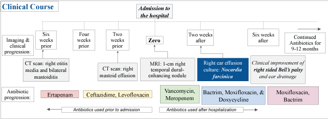 Figure 2: Clinical course and antibiotic progression for our patient.
View Figure 2
Figure 2: Clinical course and antibiotic progression for our patient.
View Figure 2
Nocardia species are opportunistic infectious agents often found in the immunocompromised population that commonly cause chronic granulomatous suppurative infections [3]. Due to their ubiquitous presence in the environment, isolation of this gram positive bacteria needs to correlate with clinical findings. Commonly, patients will be chronically immunosuppressed, either drug-induced (i.e. corticosteroids) or non-drug-induced (i.e. chronic granulomatous disease, chronic alcoholism, diabetes mellitus, Human Immunodeficiency Virus (HIV) infection) [1]. Diagnosis of this pathogen requires proof of the organism in tissue, cultures, or both. As cultures may take five or more days to identify nocardia, newer techniques, such as 16 sRNA amplification, that allow quicker identification are helpful in distinguishing nocardia species from one another to allow timely initiation of appropriate antimicrobial therapy [2].
Pulmonary nocardiosis is the most commonly encountered presentation [1], which has been reported in HIV patients [3]. Of the 67 cases of Nocardia farcinica infections reported from 2000-2012, almost 60% were found to affect the lungs [2]. Up to half of pulmonary nocardiosis cases have involvement outside the lungs, and approximately 20% of patients present only with extrapulmonary disease [1]. Common sites of these extrapulmonary manifestations include the skin, subcutaneous tissue, and the central nervous system. Rarely, bacteremia due to nocardia can occur [1]. It has been reported that Nocardia asteroides can cause mastoiditis in an HIV-infected patient [4], however, reports of N. farcinica causing disease, in particular mastoiditis, remain rare [2]. Reported cases of nocardia causing mastoiditis can be found in Table 1.
Table 1: Reported cases of Nocardia spp. mastoiditis. View Table 1
Nocardia species are known to have distinct drug susceptibility patterns. Most all species of nocardia are susceptible to TMP/SMX, and thus it has become the backbone of modern day, multi-drug treatment [1]. Nocardia farcinica has a more unique drug susceptibility pattern owing to this species being more virulent, with a mortality rate of more than 30%. Of the 67 cases reported between 2000-2012, 25 (37%) of those patients died [2]. N. farcinica has been shown to have remarkable resistance to most beta-lactams and aminoglycosides, excluding amikacin (to which all isolates have been reported as susceptible) [1,5]. Although less frequent, resistance to ciprofloxacin, imipenem, cefuroxime, and amoxicillin/clavulanic acid has been noted [5]. Additionally, meropenem has shown less efficacy against N. farcinica compared to other nocardia species [1]. In addition to TMP/SMX and amikacin, N. farcinica isolates are usually susceptible to dapsone, doxycycline, and minocycline [5]. Although resistance to ciprofloxacin occurs, the newer fluoroquinolone moxifloxacin has shown greater bactericidal activity [1]. More recently, susceptibility to oxazolidinones has been reported. While linezolid initially showed great potential in treatment of multiple nocardia species, the second-generation tedizolid has been reported as a superior and safer option, effective against N. farcinica in a case of CNS nocardiosis [5].
In our patient, broad spectrum antibiotic coverage failed to produce a clinical result, highlighting the extensive resistance of Nocardia farcinica to common antimicrobial therapy. A prolonged period of infection without clinical resolution after multiple lines of high-powered antibiotics raised suspicion for a rare organism. Pathogen identification and susceptibility testing were critical in establishing a treatment plan aimed at eradicating this infection. Due to inadequate prior treatment with IV antibiotics, the unexcluded possibility of a polymicrobial infection, and our patient's optimistic clinical picture, a multi-drug treatment plan, based on antibiotic susceptibilities, was successfully instituted.
We report a rare case of Nocardia farcinica mastoiditis in a patient with multiple myeloma and recurrent otitis media. We hope increasing awareness of this organism as a resistant pathogen will expedite diagnosis and targeted treatment in future cases.
No funding was necessary for this work.
The authors report no conflicts of interest.