Mitral valve, Perforation, Three-Dimensional transesophageal echocardiography
A 63-year-old man was referred to perform a transesophageal echocardiography (TEE) for suspicion of mitral endocarditis. The 2-Dimensional TEE showed a solution of continuity in the anterior leaflet of the mitral valve compatible with mitral perforation (Figure 1A). The Doppler color study revealed severe mitral regurgitant jet through the perforation (Figure 1B). The 3-Dimensional TEE confirmed perforation of the anterior mitral valve leaflet in the A2-A3 scallops (Figure 1C, and Figure 1D); vegetations were excluded. Perforation of mitral valve leaflets is a well known complication of infective endocartitis [1]. The incidence may be higher than whish had been reported in autopsy studies; around 8-20%. [1] 3-Dimensional TEE plays a key role in the characterization of valvular heart disease, especially, the mitral valve disease [2-4]. In spite of high quality of 2-Dimensional TEE, the 3-Dimensional TEE proved to be more accurate to characterize the localization of the anterior mitral leaflet perforation and to exclude mitral valve vegetations than the 2-Dimensional TEE [4,5]. It provide anatomical details of the mitral valve and precise localization of the perforation, which is very important for surgical planning because the en face view can simulate the view of the surgeons [6]. The present case illustrate the great accuracy of the real time 3-Dimensional TEE in the morphological analysis of mitral valve perforation.
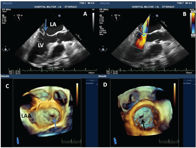
Figure 1: (A) 2-Dimensional transesophageal echocardiography revealed contiguity solution of the anterior leaflet of the mitral valve; (B) Doppler color study showing severe mitral regurgitation through the perforation; (C and D) Real time 3-Dimensional transesophageal echocardiography revealing the anterior leaflet of the mitral valve perforation in A2-A3 scallops; "en face" view from the left atrium and the left ventricle respectively. LAA - Left Atrial Apenddage; LA - left atrium; Ao - Aorta; LV - left ventricle.