
Primary cardiac lymphomas (PCL) are rare tumors with heterogeneous presentation, often difficult to diagnose, requiring a high level of clinical suspicion. An attempted diagnosis is fundamental for effective treatment. We report a very rare case of a PCL in a middle-age female patient that presented with atrial flutter, atrioventricular conduction disorder and a secondary autoimmune haemolytic anemia with cold agglutinin syndrome. The investigation was challenging and a definite diagnosis was achieved by histopathological study and corroborated by regression after chemotherapy.
Cardiac tumors, Diffuse large B-cell lymphoma, Autoimmune haemolytic anemia
AF: Atrial Fibrillation; AIHA: Autoimmune Haemolytic Anemia; AV: Atrioventricular; CAS: Cold Agglutinin Syndrome; CMR: Cardiac Magnetic Resonance; DLBCL: Diffuse Large B-Cell Lymphoma ECG: Electrocardiogram; LDH: Lactate Dehydrogenase; NHL: Non-Hodgkin Lymphoma; NT-Probnp: N-Terminal Brain Natriuretic Peptide; PCL: Primary Cardiac Lymphoma; PET: Positron Emission Tomography; R-CHOP: Rituximab, Cyclophosphamide, Doxorubicin, Vincristine And Prednisolone; SUV: Standardized Uptake Value; TACT: Thoracoabdominal Computed Tomography; TTE: Transthoracic Echocardiogram
Primary cardiac tumors are extremely rare entities, with incidence ranging from 0.001% to 0.03% and among these PCL accounts for only 1.3% [1]. They are typically non- Hodgkin lymphoma (NHL) with diffuse large B-cell lymphoma (DLBCL) being the most frequent, and are often associated with immunodeficiency states and very rarely affect immunocompetent patients [2]. They can occur as intracardiac or pericardiac masses or more rarely, infiltrate the myocardium. Clinical presentation is very heterogeneous, often with non-specific symptoms, which contributes to late diagnosis and often comprises of heart failure, pericardial effusion and more rarely atrioventricular (AV) conduction disorders. Other systemic manifestations as autoimmune haemolytic anemia (AIHA) are very rarely described. Clinical awareness and suspicion is crucial and a multimodal imaging approach is often necessary for a complete and precise diagnose.
We report the case of a middle-age female patient in her 60s that presented with dyspnoea, cough and fatigue for two weeks. One week before the patient was admitted with similar complaints and an electrocardiogram (ECG) confirmed atrial flutter with complete AV block (Figure 1). Transthoracic echocardiogram (TTE) was normal. A pacemaker DDDR was implanted and one day later the patient was discharged with anticoagulation.
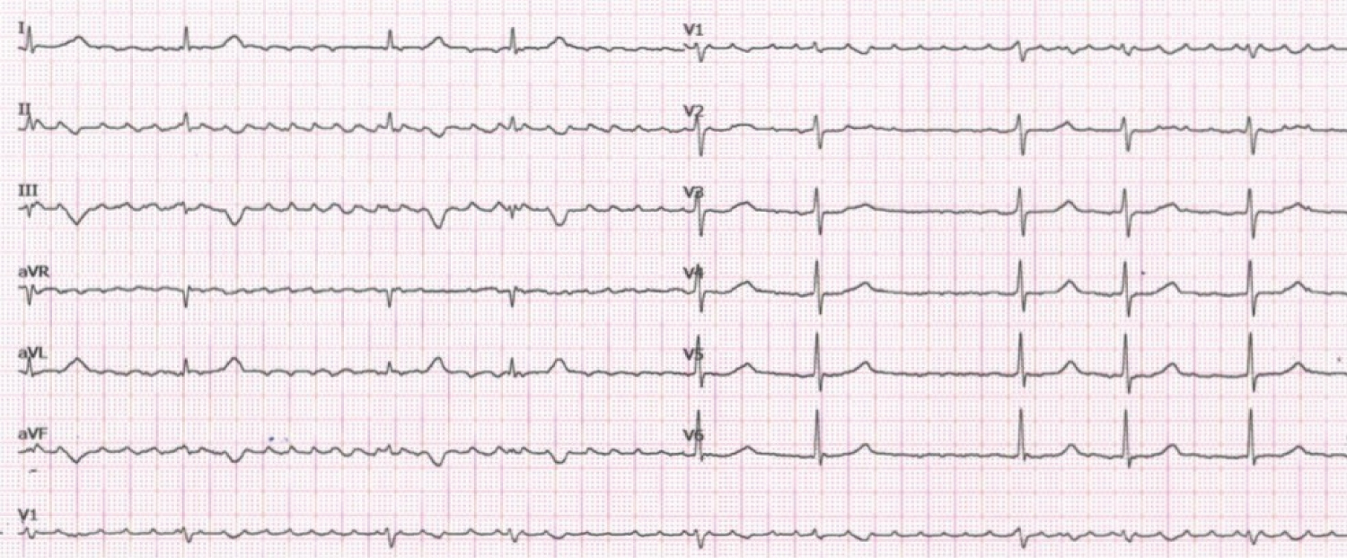 Figure 1: Electrocardiogram (ECG). 12-lead ECG showing atrial flutter with complete atrioventricular block.
View Figure 1
Figure 1: Electrocardiogram (ECG). 12-lead ECG showing atrial flutter with complete atrioventricular block.
View Figure 1
On examination the patient was afebrile, heart rate 56 bpm, blood pressure 110/67 mmHg, SpO2 98% in room air. No lymphadenopathy or peripheral oedema was noted. Cardiac auscultation was normal and pulmonary auscultation revealed bilateral basal crackles and the remaining examination was normal.
The patient had history of breast ductal carcinoma (T2N1M0) 28 years ago that was treated with mastectomy, adjuvant chemotherapy (methotrexate, mitoxantrone and 5-fluorouracil) and hormonal therapy (goserelin and tamoxifen during 5 years) with regular follow-up in Oncology with annual mammography and no other relevant past medical or family history. She was medicated with apixaban since the last week.
Laboratory test revealed aggravated normocytic normochromic anaemia (Hb 8.4 g/dL, Htc 23.3%, previous values from one week earlier were Hb 10.2 g/dL, Htc 30.9%), mild leukocytosis (11.090/uL) and elevated N-terminal brain natriuretic peptide (NT-proBNP) 50001 pg/mL. ECG revealed atrial fibrillation (AF). TTE normal biventricular function and a small circumferential pericardial effusion (15 mm) without haemodynamic compromise. Chest radiography was normal. Pacemaker interrogation showed AF and normal parameters. A diagnosis of congestive heart failure was assumed and she was hospitalized for further investigation and therapy optimization. One day later she had clinical deterioration with one auto-limited episode of hypotension (BP 82/58 mmHg) and abdominal diffuse pain. TTE excluded cardiac tamponade. Laboratory results revealed leukocytosis (21.400/uL) and C-reactive protein (11 mg/dl). Considering the hypothesis of a systemic infection, blood cultures were collected and large spectrum antibiotic therapy was initiated. A thoracoabdominal computed tomography (TACT) showed an irregular pseudonodular mass in the inferior interventricular sulcus, pericardial effusion and several enlarged mediastinal lymph nodes. Considering the recent pacemaker implantation, the hypothesis of a lead complication was contemplated and the pacemaker was removed. The patient remained in AF with preserved AV conduction. Later she continued to deteriorate with jaundice and increasing abdominal pain and anaemia was worse despite transfusion of two red blood cell units (Hb 6.8 g/dL) with increased lactate dehydrogenase (LDH) (914 U/L) and bilirubin (total 4.8 mg/dl, direct 1.5 mg/dl). An AIHA was suspected and a direct antiglobulin test was positive for IgM (3+), C3c (4+) and C3d (4+) confirming the diagnosis of a cold agglutinin syndrome (CAS). Treatment with prednisolone and rituximab was initiated with mild clinical improvement. Three days later a cardiac magnetic resonance (CMR) confirmed the diagnosis of an infiltrative mass in the inferior interventricular sulcus with 20 × 40 mm in basal short axis isointense on T1-weighted images, hyperintense on T2-weighted images, with heterogeneous late gadolinium enhancement (Figure 2) and peripheral enhancement with central hypoperfusion suggesting a malignant aetiology. Left and right ventricular function was normal with hypokinesia of the right ventricular inferior wall. A positron emission tomography (PET) scan showed a conglomerate of lymph nodes in the aortopulmonary region (standardized uptake value (SUV)) of 11.4 and in the basal portion of the inferior wall and interventricular septum (SUV 8.3).
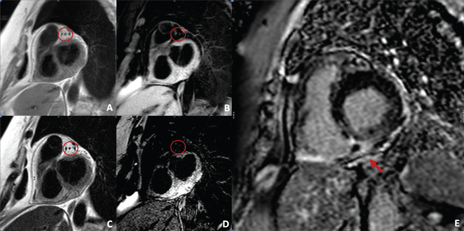 Figure 2: Cardiac magnetic resonance (CMR) imaging. CMR short-axis shows an infiltrative mass in the inferior interventricular sulcus, isointense on T1-weighted images (A and B, without and with fat suppression) and hyperintense on T2-weighted images (C and D, without and with fat suppression). Short-axis late gadolinium enhancement image showing heterogenous late enhancement (E).
View Figure 2
Figure 2: Cardiac magnetic resonance (CMR) imaging. CMR short-axis shows an infiltrative mass in the inferior interventricular sulcus, isointense on T1-weighted images (A and B, without and with fat suppression) and hyperintense on T2-weighted images (C and D, without and with fat suppression). Short-axis late gadolinium enhancement image showing heterogenous late enhancement (E).
View Figure 2
After discussion in a multidisciplinary team with Cardiologists, Haematologists and Cardiac Surgeons a surgical biopsy through sternotomy was performed. On surgical inspection an infiltrative mass on the inferior interventricular sulcus composed of four infracentimetric white nodules was identified and five biopsies were obtained. Immunohistochemical findings revealed a DLBCL non germinal center type, CD45+, CD20+, BCL2+, BCL6+, MUM1+, Ki67 95%.
The patient was transferred to an Oncology Center and chemotherapy was initiated according to established protocols with rituximab, cyclophosphamide, doxorubicin, vincristine and prednisolone (R-CHOP). Six cycles of R-CHOP were administrated with complete response and progressive clinical improvement. A follow-up CMR was performed with complete regression of the DLBCL. To the date the patient is asymptomatic with no signs of heart failure or other complications and remains in AF with preserved AV conduction and did not require a pacemaker.
We present a very rare case of a PCL that presented with atrial flutter and AV conduction disorder and a secondary cold agglutinin haemolytic anaemia.
Our patient had a very uncommon presentation of a PCL with pericardial effusion, atrial flutter with complete AV block and a secondary CAS.
Complete AV block often requires permanent pacing, unless a reversible cause can be identified. In this case AV block was initially thought to be primary, probably due to idiopathic fibrosis and sclerosis, since this is the most common aetiology and a pacemaker DDDR was implanted. A secondary cause for complete AV block was only suspected when multimodality imaging detected an infiltrative cardiac mass.
Considering the short time after pacemaker implantation the pericardial effusion and the elevated inflammatory parameters were assumed to be a complication of its implantation and the decision for removing it was made and antibiotic therapy was initiated. Since then the patient remained with preserved AV conduction and did not require a definite pacemaker. Complete AV block related to PCL can be transient and can resolve after effective treatment of the tumor and so, it may be reasonable to delay pacemaker implantation until after treatment.
A recurrence or metastasis of breast cancer was hypothesized as a cause for a cardiac mass but was unlikely considering that the patient had regular follow-up in Oncology with a recent normal mammography and the TACT showed no related abnormalities. The patient had no obvious risk factors for lymphoproliferative disease except for chemotherapy treatment in the past, but it was over 28 years ago and so this hypothesis seemed unlikely.
According to autoantibodies cold type AIHA is the rarest form and CAS can be secondary to infections, autoimmune or lymphoproliferative disorders. CAS secondary to DLBCL has been reported but is very rare [3]. In our case AIHA was initially thought to be secondary to a systemic infection, to recurrence of breast cancer or, as it turned out to be, secondary to a lymphoproliferative disorder.
The work-up was complex and clinician awareness was essential. After suspicion of a cardiac mass diagnostic work-up involved multimodality noninvasive imaging. TTE is widely available and allowed diagnose of pericardial effusion. TACT was useful in identifying the mass and lymphadenopathies. CMR was essential to access the characteristics of the mass and its relationship to surrounding structures predicting malignancy, and plays a role in monitoring treatment response. PET helped to evaluate the nature of the mass and exclude dissemination. Histopathological study is the gold standard and allowed definite diagnosis and the determination of the adequate treatment. In conclusion the investigation was challenging and a multidisciplinary team approach was fundamental in decision making and orientation.
The authors have nothing to disclose.
Self-finance.