Idiopathic root resorption of the permanent dentition is a very rare condition which can be diagnosed during routine dental examinations. A 13-year-old German female was referred to the Unit of Pediatric Dentistry regarding fistulas of all her 1st permanent molars as the patient did not have any specific symptoms with regard to her teeth. All the first molars have been affected in a symmetrical pattern due to a massive idiopathic external root resorption. Clinically, the oral hygiene was very good, the teeth were sealed and no dental caries. A histological and hematological examination has been conducted. The past medical history revealed that she had a congenital kidney anomaly and she had spent almost the first six months of her life in the hospital. Since that time she has been regularly visiting the nephrology department. There was neither a family history regarding the root resorption nor the congenital kidney anomalies.
Idiopathic external root resorption is a very rare condition. It has been reported in single and multiple teeth. Pathological root resorption is related to local factors and sometimes related to systemic factors, L Khojastepour, et al. Several local causes that may lead to pathological root resorption have been discussed: Orthodontic therapy, dental trauma, periapical or periodontal inflammation, tumors, cysts, occlusal stress, impacted and supernumerary teeth, transplantation and reimplantation. L. Khojastepour, et al., The systemic causes that have been implicated are hyperparathyroidism, hyperparathyroidism, hypophosphatemia, hyperphosphatemia, Gaucher' s disease, Paget' s disease of the bone, Goltz syndrome, Papillon-Lefevre syndrome, anachoresis, Turner syndrome as well as the endocrine disturbances.
"Idiopathic" is a term which means that the main etiological factor for root resorption cannot be identified. Two types of idiopathic root resorption have been observed, apical and cervical. Cervical root resorption starts cervically and progresses towards the pulp. Apical root resorption starts apically and progresses towards the coronal area which leads to a short and round remaining root. In a recent literature review Cholia, et al. [1] the idiopathic apical root resorptions were found to be more common in the upper jaw and molar region than in the lower jaw and single root teeth; however, root resorption may affect only the 1st molars in all quadrants in a symmetrical pattern. This article describes a case of idiopathic apical root resorption in which no local cause could be identified. A history of a congenital kidney disease could be implicated as a systemic cause.
A 13-year-old German female was referred to the Unit of Pediatric Dentistry regarding fistulas of all her 1st permanent molars as the patient did not have any specific symptoms with regard to her teeth. The past medical history revealed, that she had a congenital kidney anomaly and she had spent almost the first six months of her life in the hospital. Since that time she has been regularly visiting the nephrology department. There was neither a family history regarding the root resorption nor the congenital kidney anomalies. In our first intra-oral examination the patient revealed a caries-free dentition (sealants on molars with the resin-based dental material, no initial lesions) and very good oral hygiene, with healthy gingiva. There was no history of dental trauma and no history of orthodontic treatment but an orthodontic treatment plan.
All affected teeth (1st molar in each quadrant) had been extracted after injection of local anesthesia. The infected tissues had been debrided after extraction. A biopsy from the infected tissue has been taken and has been sent immediately to the pathologist (Figure 1, Figure 2, Figure 3 and Figure 4).
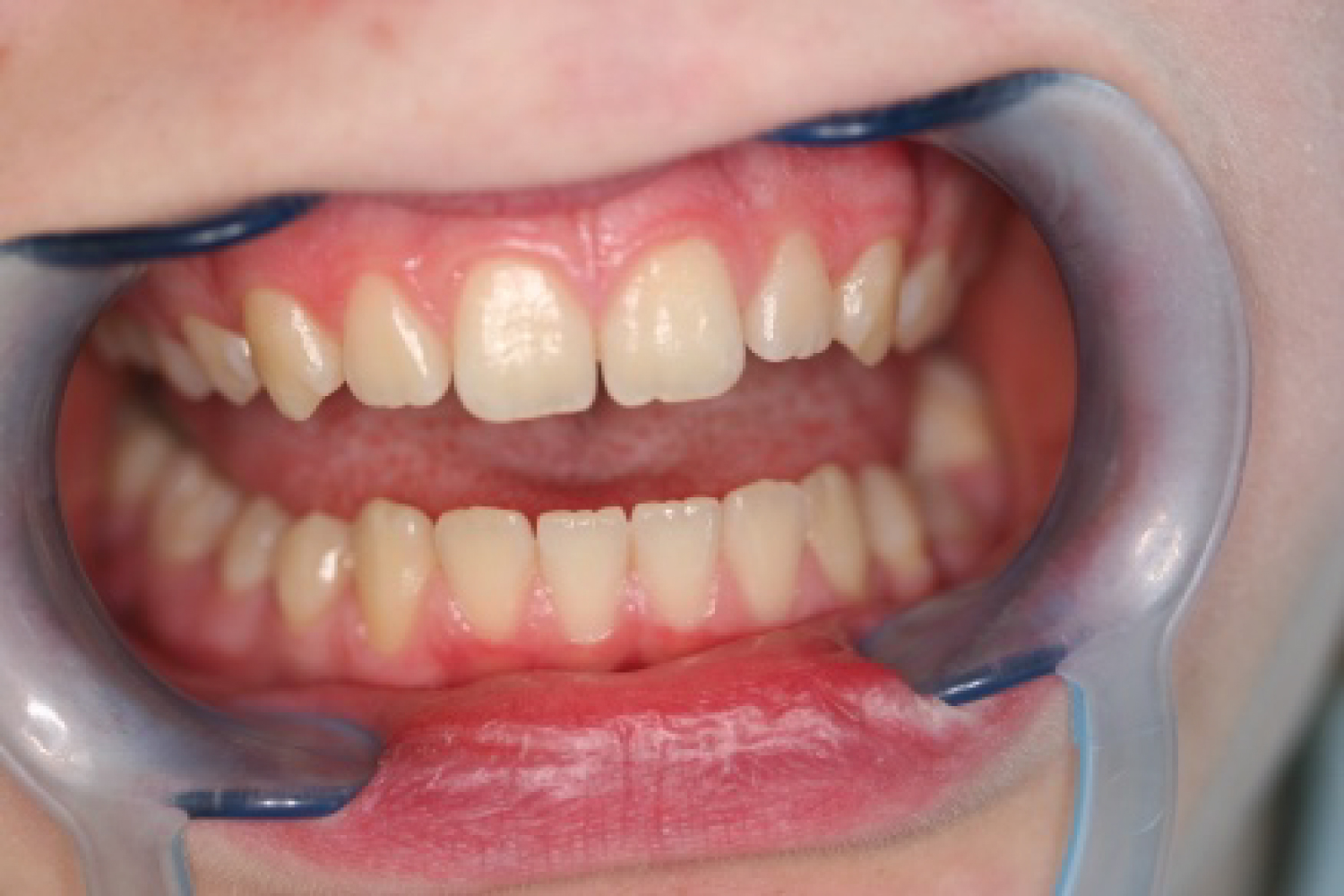 Figure 1: Anterior aspect.
View Figure 1
Figure 1: Anterior aspect.
View Figure 1
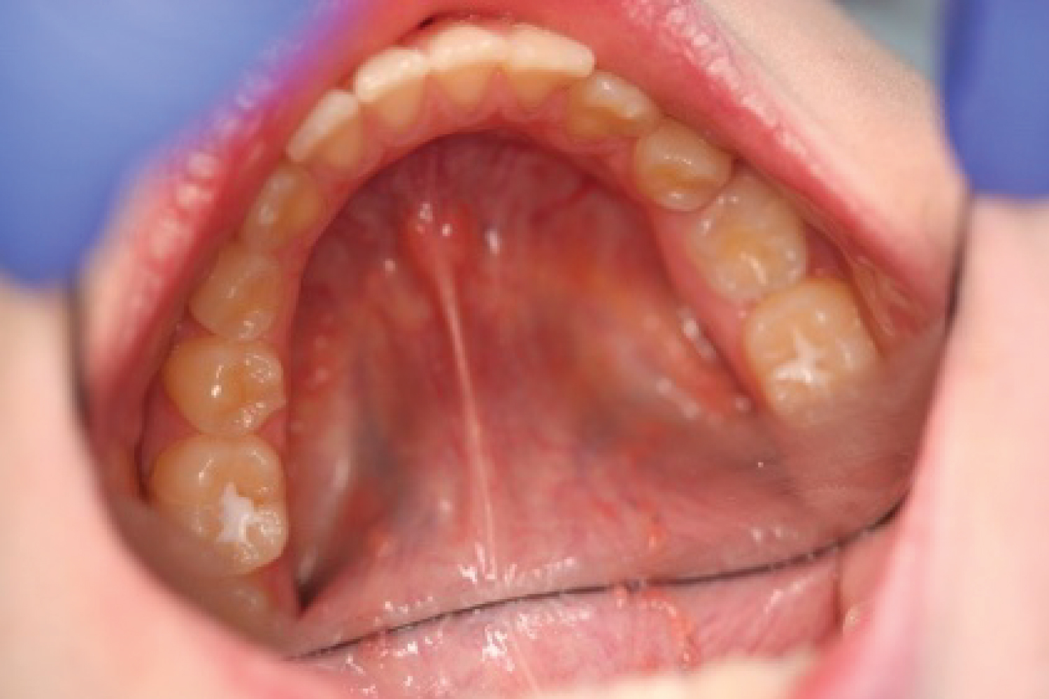 Figure 2: Lower arch with sealed teeth.
View Figure 2
Figure 2: Lower arch with sealed teeth.
View Figure 2
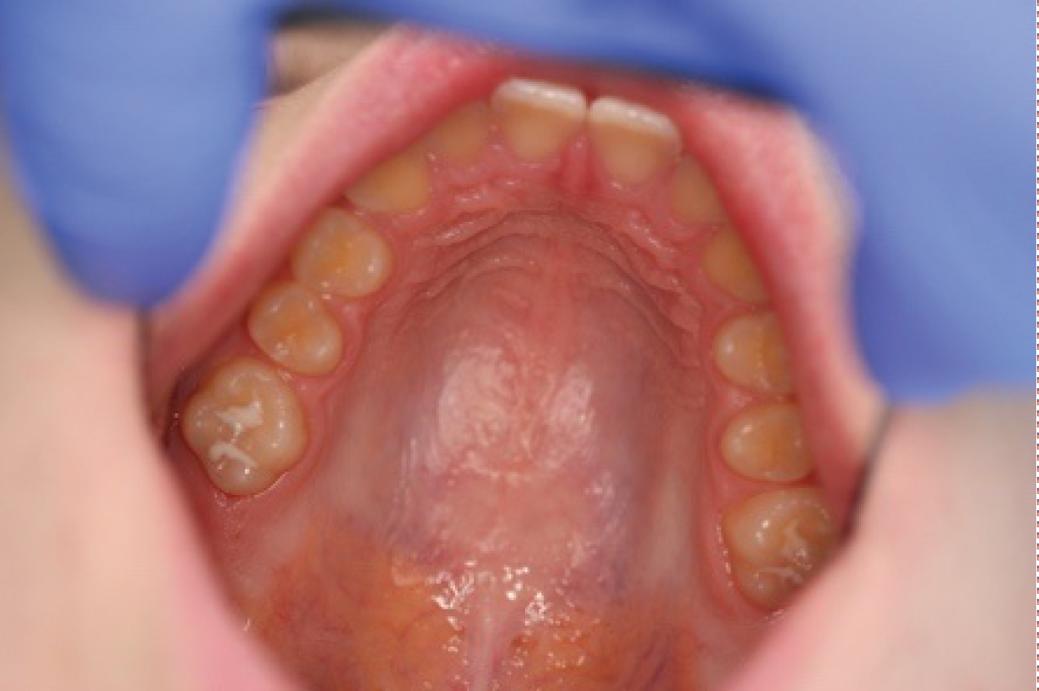 Figure 3: Upper arch with sealed teeth.
View Figure 3
Figure 3: Upper arch with sealed teeth.
View Figure 3
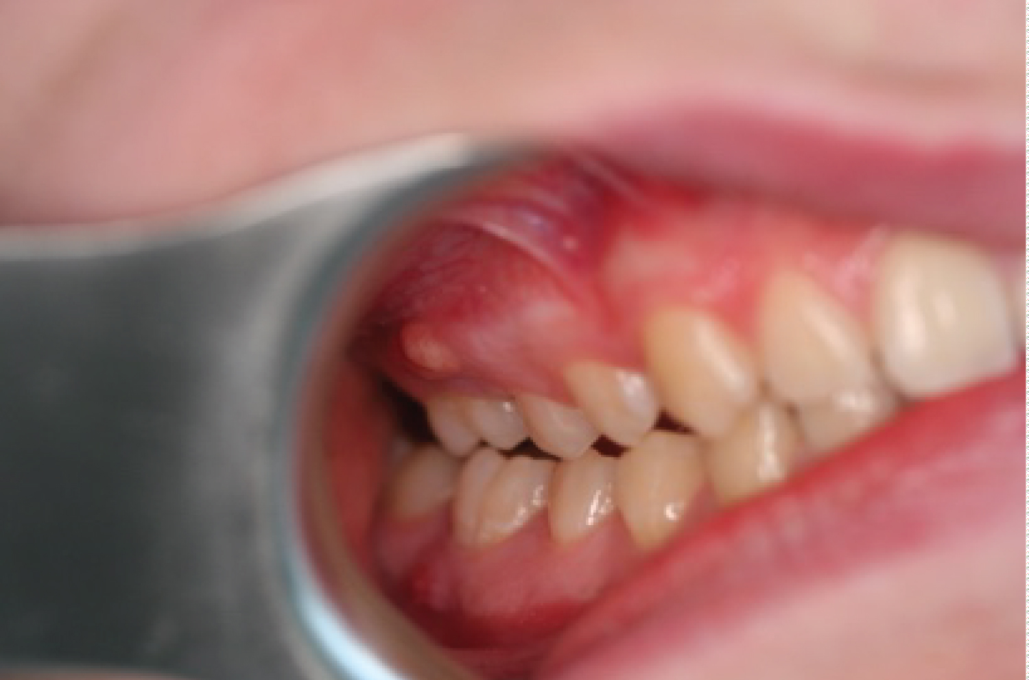 Figure 4: Fistula related to all 1st molars #16.
View Figure 4
Figure 4: Fistula related to all 1st molars #16.
View Figure 4
A panoramic radiograph revealed severe apical root resorptions affecting all maxillary and mandibular first molars.
The severity of the resorption increased in the cervical area of the lower first molars. This had progressed almost to the radicular furcation, but there was no evidence of alveolar bone loss or periradicular periodontitis. The hematological and biochemical screening was within the normal range. A diagnosis of multiple idiopathic external apical root resorption (MIEARR) was mostly based on the radiographic findings (Figure 5).
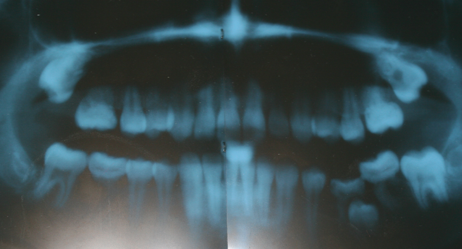 Figure 5: Panorama for whole teeth.
View Figure 4B
Figure 5: Panorama for whole teeth.
View Figure 4B
Periapical X-ray shows an obvious radiolucency related to the deformed roots (Figure 6).
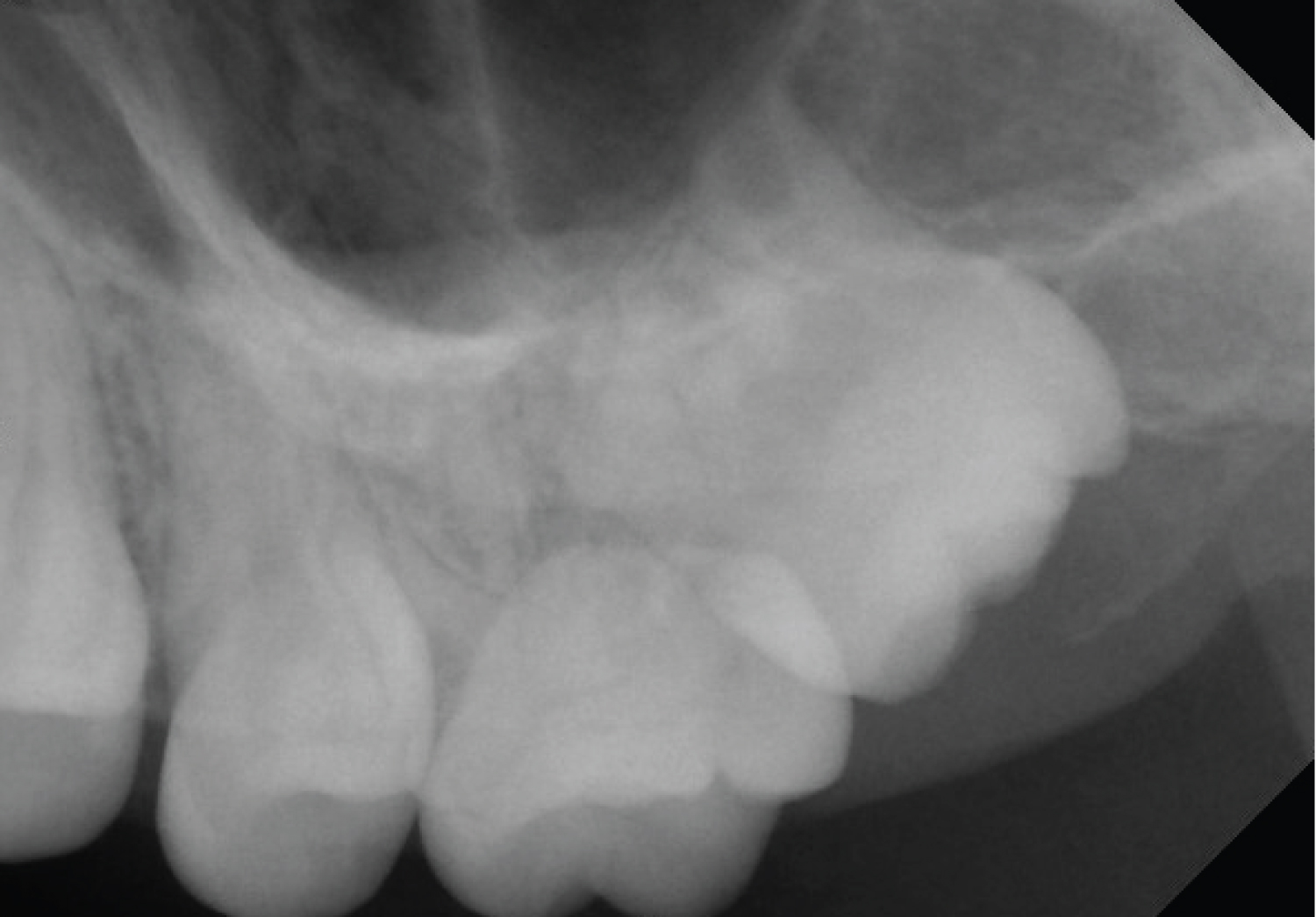 Figure 6: Shows x-rays for upper 1st molar (1#6)
View Figure 6
Figure 6: Shows x-rays for upper 1st molar (1#6)
View Figure 6
An extracted upper 1st molar (1#6) shows a hole due to the massive resorption in the coronal part of the root. Granulation tissue has been seen on the coronal part of the root surface (Figure 7).
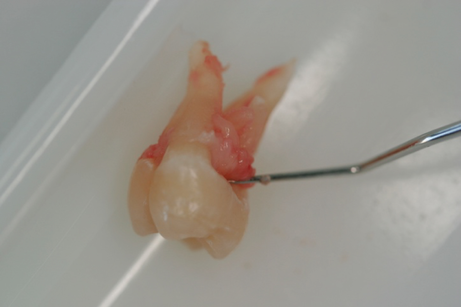 Figure 7: Shows an extracted upper 1st molar (1#6).
View Figure 4B
Figure 7: Shows an extracted upper 1st molar (1#6).
View Figure 4B
Periapical X-ray shows an obvious radiolucency related to the deformed roots (Figure 8).
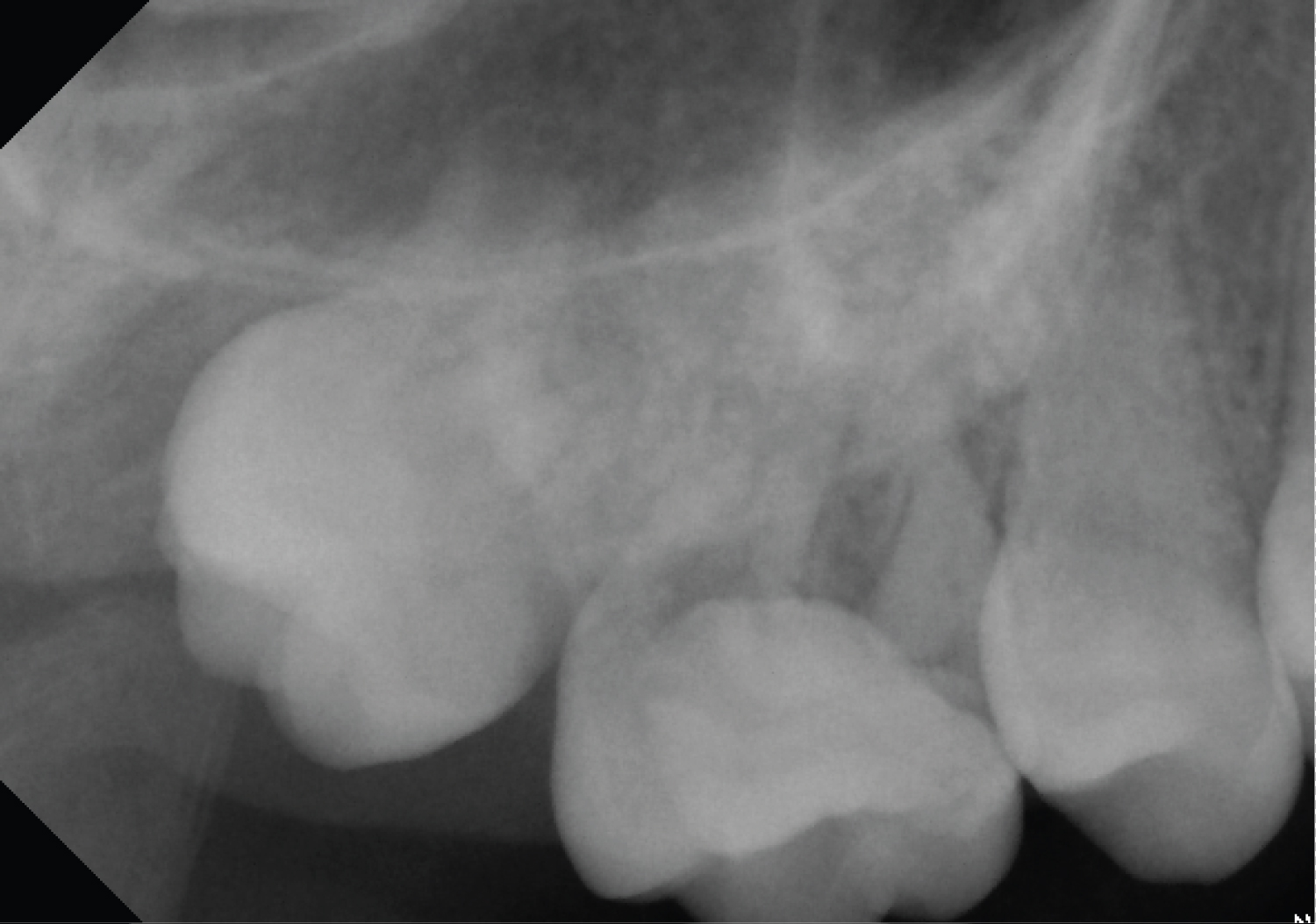 Figure 8: Shows x-rays for upper 1st molar (2#6).
View Figure 4B
Figure 8: Shows x-rays for upper 1st molar (2#6).
View Figure 4B
An extracted upper 1st molar (2#6) shows deformed roots. The apical part of the roots was smooth and rounded with infected tissue on the remaining surface of the deformed roots (Figure 9).
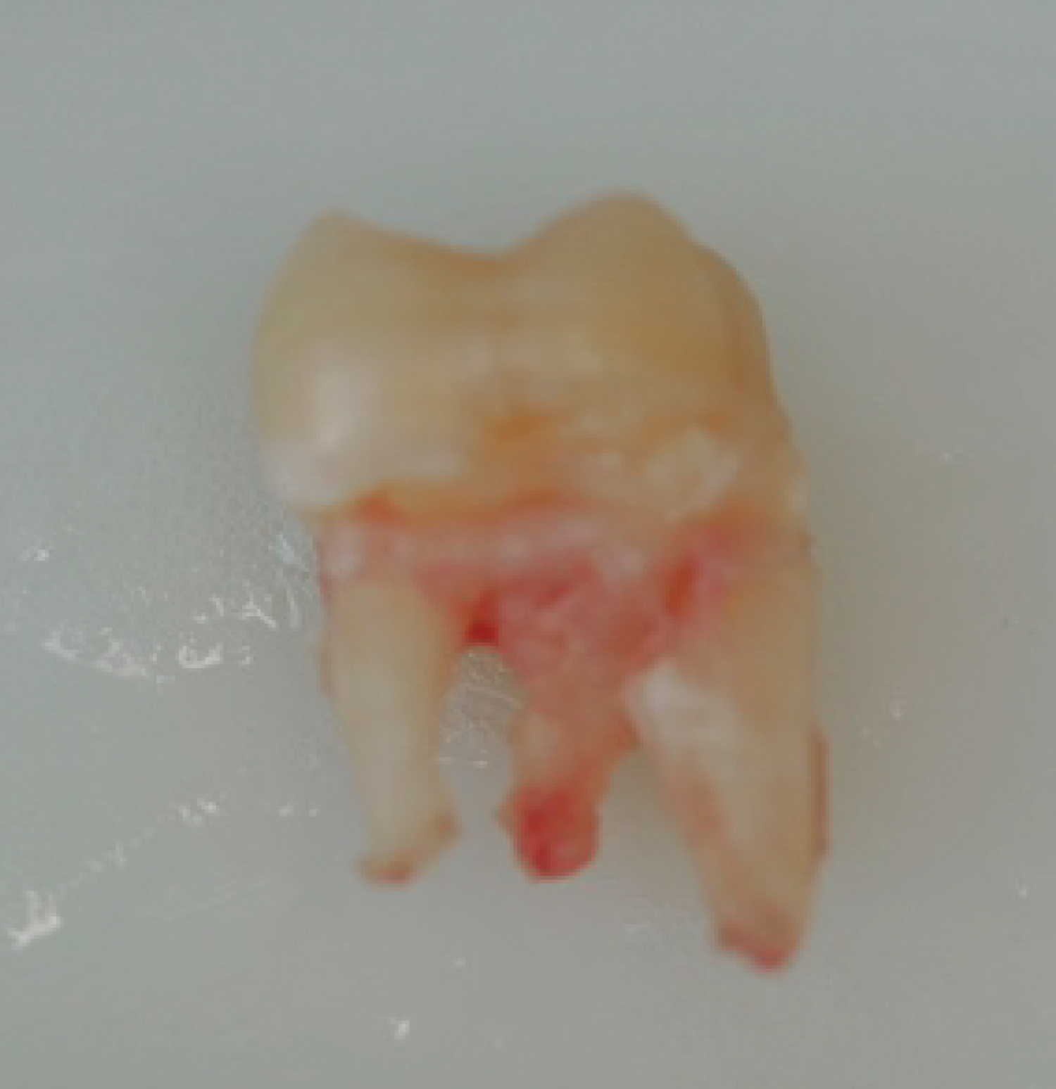 Figure 9: Shows an extracted upper 1st molar (2#6).
View Figure 4B
Figure 9: Shows an extracted upper 1st molar (2#6).
View Figure 4B
Periapical X-ray shows an obvious radiolucency related to the deformed roots (Figure 10).
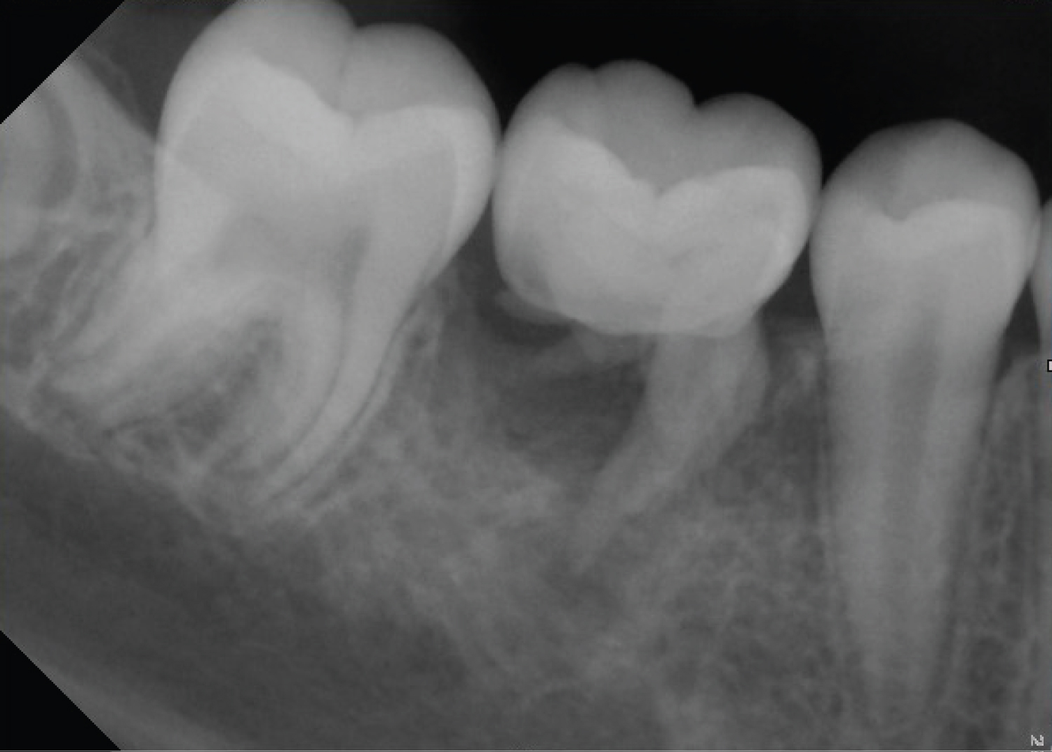 Figure 10: Shows x-rays for upper 1st molar (3#6).
View Figure 4B
Figure 10: Shows x-rays for upper 1st molar (3#6).
View Figure 4B
An extracted lower 1st molar (3#6) shows deformed roots. The surface of the apical part was smooth and rounded with infected tissue on the remaining surface of the deformed roots (Figure 11).
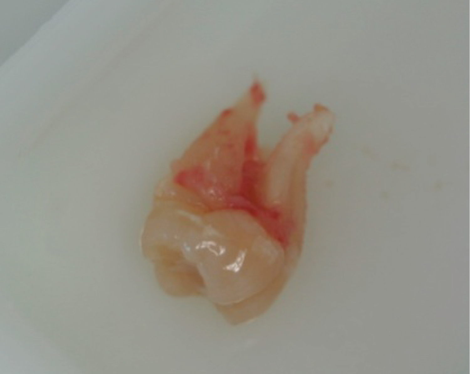 Figure 11: Shows an extracted upper 1st molar (3#6).
View Figure 4B
Figure 11: Shows an extracted upper 1st molar (3#6).
View Figure 4B
An extracted lower 1st molar (4#6) shows deformed roots. A granulation tissue has been seen on the root surfaces. The distal root is shorter than the mesial one (Figure 12).
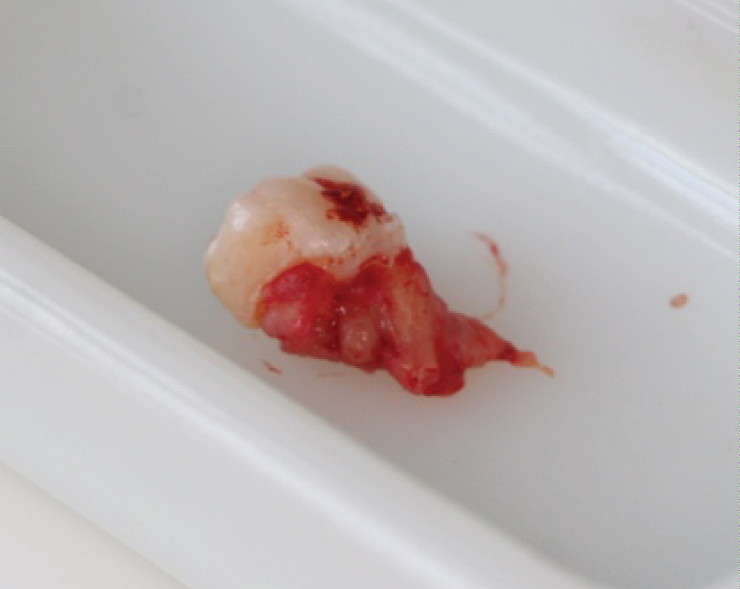 Figure 12: Shows an extracted upper 1st molar (4#6).
View Figure 4B
Figure 12: Shows an extracted upper 1st molar (4#6).
View Figure 4B
Few cases of multiple idiopathic apical root resorption (MIARR) exist in the literature. The first well-documented report was in 1930 [2] and since then more cases were presented [3-5]. With no absolute etiological factor identified we considered this case as multiple idiopathic apical root resorptions. All teeth had vital pulps and there was no periodontal or periapical inflammation. Resorption was found incidentally in the panoramic view and the patient was totally asymptomatic. No local etiologic factor was detected and clinical appearance of the teeth and periodontium were normal.
Regarding the number of affected teeth in literature review which were about eighteen on average, [4], in this report only three teeth were involved. It may be related to the age of the patient compared with other reported cases. The average reported age was 23.2 years [4]. The present case was among a few cases who were under 21-years-old at the time of detection. The condition has been reported to have a predilection for young females [6] and 14 cases of MIARR have been being presented in females till now [7-15].
The etiology of multiple idiopathic external apical root resorptions still needs further investigations to reveal its molecular mechanism. So far, only symptom-based treatment is advisable.