Hypertrophic pyloric stenosis (HPS) is a disorder of infants that results in an abnormal thickening of the pyloric sphincter. Surgical division of the pyloric muscle fibers under general anesthesia is the standard treatment. There is currently no realistic simulation model of hypertrophic pyloric stenosis. Surgeons learn the procedure on live infants. Our objective was to determine if an adult embalmed cadaver pylorus, a readily available resource at most medical schools, could serve as a simulation model of HPS for the performance of an open infant pyloromyotomy.
A general surgeon, with training in pediatric surgery and surgical experience with pyloromyotomy, performed pyloromyotomies on all embalmed cadavers in a medical school anatomy lab, utilizing an anterior longitudinal incision and spreading of the pyloric fibers with a small curved hemostat. The surgeon rated each cadaver pylorus and pyloromyotomy as: 1) Realistic simulation of HPS and pyloromyotomy, or 2) Poor simulation of HPS or pyloromyotomy. Following pyloromyotomy, the surgeon measured the thickness of the pylorus. The surgeon was blinded to the age, gender and cause of death of the cadaver.
Based on expert opinion, 23/30 (77%) of cadavers provided realistic simulation of HPS and pyloromyotomy. Pyloric thickness ranged from 1-7 mm. A pyloric thickness of 3 mm or greater provided the best simulation experience.
Based on one surgical expert's opinion, the pylorus of adult embalmed cadavers could provide a valuable open or laparoscopic simulation training experience for surgeons.
Hypertrophic pyloric stenosis, Pylorus, Pyloromyotomy, Cadaver simulation
Hypertrophic pyloric stenosis (HPS) is the abnormal thickening of the muscular layer of the pyloric sphincter. Never present at birth, this potentially fatal condition presents in otherwise healthy infants, typically 2 to 8 weeks of age. Male to female ratio is 4:1 [1]. The primary function of the pylorus is to regulate gastric emptying. Hypertrophy of the circular muscle of the pylorus results in constriction and obstruction of the gastric outlet leading to non-bilious projectile vomiting and accompanying fluid, acid base and electrolyte abnormalities. The exact cause of HPS is unknown but a lack of nitric oxide synthase, a mediator of smooth muscle relaxation within the digestive tract, has been implicated, as well as various genetic factors (as evidenced by family history, and race, Caucasian) and environmental factors (as evidenced by seasonal variability, and increased incidence with transpyloric feeding in premature infants) [1,2]. A thorough history and physical exam will commonly be highly suggestive of HPS. Visible gastric peristalsis may be observed on the abdominal wall and palpation of an "olive" tumor in the epigastrium is pathognomonic for HPS. Experts consider the ultrasound measurement of 3 mm or greater or a pyloric length of greater than 15-18 mm to be diagnostic of HPS. When equivocal, an upper gastrointestinal contrast study may be required. [3]. The mainstay of treatment of HPS is pyloromyotomy, first described by Ramstedt in 1912, which surgeons now perform utilizing an open or laparoscopic technique [4-6]. The training of residents, fellows, and surgeons in the technique of pyloromyotomy currently requires live patients [7]. The only pyloromyotomy simulation model that has been described utilizes an inanimate constructed object [8]. Since both laparoscopic and open techniques are currently utilized throughout the United States and neither has been identified to be superior [9] a training platform that simulates an open pyloromyotomy procedure is pertinent. This report is the first to describe the use of the embalmed adult cadaver pylorus as a training platform for pyloromyotomy.
Our medical school anatomy lab purchases approximately thirty-two embalmed cadavers on a yearly basis for anatomical dissection by our medical students. A single general surgeon, with fellowship training in pediatric surgery and surgical experience with pyloromyotomy, performed pyloromyotomies on all embalmed cadavers present in a medical school anatomy lab. The surgeon conducted the procedure in the usual fashion, utilizing an anterior longitudinal incision into the pyloric serosa which was then deepened with the blunt end of a scalpel handle. The surgeon then divided the remaining circular muscle fibers of the pylorus by spreading with a small curved hemostat (Figure 1). The surgeon rated each pyloromyotomy according to how closely the sensation of performing the cadaver pyloromyotomy simulated infantile HPS and pyloromyotomy: 1) Realistic simulation of HPS and pyloromyotomy, or 2) Poor simulation of HPS or pyloromyotomy. Following the cadaver pyloromyotomy, the surgeon measured the pyloric muscle sphincter. The pyloric muscle thickness and length were measured with a straight ruler. The thickest portion of the pylorus, was invariably found to be in the mid portion, as is similarly found in infants. The surgeon was blinded to the cadaver age, gender and cause of death. Thirty of thirty-two cadavers were found to have a pylorus accessible for evaluation. Two cadavers had an inaccessible pylorus due to disease (extensive peritoneal metastatic disease) and surgery.
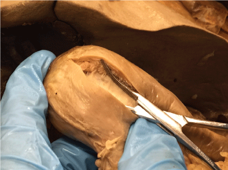 Figure 1: Cadaver pyloromyotomy procedure.
View Figure 1
Figure 1: Cadaver pyloromyotomy procedure.
View Figure 1
The pylorus of twenty-three of the thirty cadavers (77%) provided realistic simulation of an open infant pyloromyotomy. 23/23 (96%) of the cadavers that had a pyloric thickness of 3 mm or greater provided realistic simulation (Table 1). There was no apparent disruption of the duodenal mucosa during any of the procedures. Two of the 32 cadavers were not included in the evaluation because the pylorus was not accessible due to existing abdominal disease or surgery. Pyloric thickness ranged from 1-7 mm. The mean thickness for female cadavers was 4.25 mm and for male cadavers was 4.0 mm. There is no significant difference between males and females in terms of the mean and median thickness of cadaver pylorus (p-value of 0.68). In addition, there was no correlation between age of death and pyloric thickness (Pearson's correlation coefficient is 0.1732 and the estimated Spearman's correlation coefficient is 0.1195).
Table 1: Cadaver pyloromyotomy data. View Table 1
What initially drew our attention to the possibility of an adult cadaver pylorus serving as a training platform for an infantile pyloromyotomy was an anatomic finding that was identified during the usual medical student dissection of the abdomen. In a living adult the location and palpation of the pylorus can be very difficult at times. In the anatomy lab we noted that on palpation the embalmed adult cadaver pylorus was very prominent and firm in nearly all cadavers, similar to the feel of a hypertrophic pylorus in an infant.
Our cadavers are referred to as "embalmed cadavers". Embalming is a chemical process utilized to forestall decomposition and reduce the risk of infection to those who are handling the cadavers. For anatomical donors the process begins at the funeral home where 2-3 gallons of embalming solution is injected into the body. Funeral home embalming is intended to preserve the body for only a short time. The embalming process is then repeated by the anatomical donor organization embalmers utilizing six gallons of embalming solution pumped into the vascular system, under pressure and usually through the carotid artery, and then drained through the jugular or femoral veins. The embalming fluid utilized in our cadavers included a mixture of formalin, phenol, ethyl alcohol, and a mold inhibitor. There was no aspiration or injection of embalming fluid into the peritoneal cavity. The donor embalming, utilizing higher injection pressure and higher volume, is intended to preserve the body for two years.
A single surgeon, with training and experience in both open and laparoscopic pyloromyotomy, was available to conduct this experiment. Based on that surgeon's experience a binary system of evaluating the cadaver pyloromyotomy procedure was utilized. In the opinion of the surgeon, the "feel" or palpable sensation of the pyloromyotomy procedure either provided a realistic simulation of a live infant pyloromyotomy or it did not.
The "feel" or sensation of performing an open pyloromyotomy is quite unique. Generally there are three steps and each step produces a different tactile sensation. The initial step is the longitudinal division of the pyloric serosa. Our surgeon was easily able to identify the proximal and distal extents of the pylorus by palpation. Although not critical to the procedure, the pre-pyloric Vein of Mayo, sometimes used as a proximal landmark, was not clearly identifiable in the cadavers. The sensation of the serosal incision was found to be very similar to that of a living patient. The second and third steps involve division of the pyloric muscle fibers and include deepening of the serosal incision into the pyloric muscle using the blunt end of a scapel handle and then completing the pyloromyotomy by spreading and fracturing the pyloric muscle fibers with a curved clamp (Figure 1). In 75% of the cadavers the critical characteristic sensation of snapping and fracturing muscle fibers was present. Additionally, at the completion of the pyloromyotomy, the duodenal mucosa could be clearly identified, similar to a living patient (Figure 1). We then rocked both portions of the pylorus in a longitudinal fashion to confirm complete division of the pylorus. The surgeon then also compressed the stomach and duodenum to confirm no duodenal leak. Both maneuvers were conducted as would be in a live patient and both were performed successfully on all cadavers.
Bleeding is not a problem encountered during a live pyloromyotomy and the live tissues rarely require any type of hemostatic intervention, such as cautery. Similarly, there was no seepage of fluid from the pylorus noted during our procedure.
There is currently no inexpensive, easily accessible and realistic simulation model for HPS and pyloromyotomy. Surgeons train on live infants. Based on one expert's opinion in a medical school cadaver lab, surprisingly, the pylorus of adult embalmed cadavers could provide a valuable simulation training experience for surgeons and surgeons in training. Since laparoscopic pyloromyotomy is now a popular technique, and laparoscopic procedures have been performed on cadavers [10], training in pyloromyotomy could be done on an embalmed adult cadaver utilizing the open or laparoscopic technique. Medical students do not routinely destroy the pylorus during dissection; therefore, the pylorus will likely be available for pyloromyotomy training without disruption of the normal anatomical training of medical students. The wide availability of cadavers at medical schools could provide safe and realistic simulation of an infant pyloromyotomy.
Thanks to Jana Schellinger, Medical School Librarian, for providing writing assistance, editing and proofreading of the article.
None.