A 72-year-old woman with metastatic carcinoid/neuroendocrine tumour NETG1 presented with progressive right heart failure and recurrent marked ascites. An echocardiogram showed a significantly dilated right ventricle with moderate systolic impairment. The tricuspid valve leaflets were tethered resulting in poor coaptation causing severe tricuspid regurgitation (Panel A). It was felt that the TR was a result of carcinoid affecting the tricuspid valve. Therefore cardiac magnetic resonance imaging (CMR) was performed to quantify RV function. CMR (Panel B) confirmed severe tricuspid regurgitation with a regurgitant volume of 63ml and regurgitation fraction of 48%. The right ventricle was severely dilated with preserved systolic function. The tricuspid valve leaflets were thickened but significantly tented and restricted. There was apical displacement (1.1cm/ m2) of the septal leaflet and the question was raised whether it was an Ebistenoid valveTransoesophageal echocardiography (Panel C) confirmed apical displacement of the septal leaflet suggestive of Ebstein's anomaly. The origin of the tricuspid regurgitant jet also appeared to be from an apical position (Panel D). At surgery confirmed an Ebstein component to the tricuspid valve with atrialisation of the posterior right ventricle. The tricuspid valve appearances were typical for carcinoid with plaques and contraction of the leaflet tissue. She underwent tricuspid valve replacement with size 29 St Jude mechanical valve and recovered well.
The most common cause of tricuspid regurgitation is not a primary tricuspid valve disease but rather an impaired valve coaptation caused by a dilation of the right ventricle and/or of the tricuspid annulus Primary tricuspid valve disease causing tricuspid regurgitation in adults is uncommon. Ebstein's anomaly is the most common congenital heart disease affecting the tricuspid valve. Carcinoid heart disease causes thickening and retraction of the tricuspid valve leaflets resulting in tricuspid regurgitation. To our knowledge, this is the first reported case of carcinoid heart disease in Ebstein's anomaly.
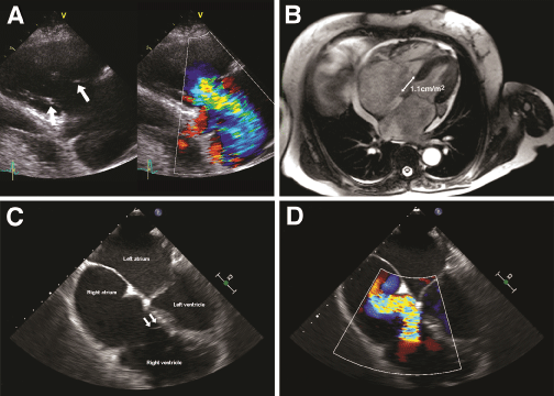
Panel A: On the parasternal RV inflow view, the tricuspid valve leaflets were severely tethered (arrows) with poor coaptation resulting severe/free tricuspid regurgitation.
Panel B: Cardiac magnetic resonance revealed a severely dilated right ventricle with preserved function. There was apical displacement of tricuspid valve septal leaflet with a separation of mitral and tricuspid planes of 1.1cm/m2.
Panel C: There was marked apical displacement of the tricuspid valve septal leaflet insertion (arrows) demonstrated on transoesophageal 4-chamber view at the mid oesophageal level.
Panel D: The origin of the tricuspid regurgitant jet appears to originate from an apical position on colour-Doppler imaging.