IMAGE ARTICLE | VOLUME 1, ISSUE 1 | OPEN ACCESS DOI: 10.23937/2474-3682/1510008
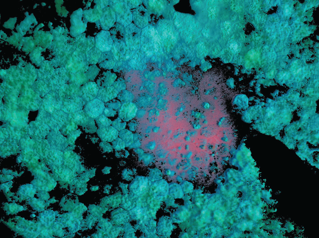
Sub-RPE Desposits - Ex Vivo Hyperspectral Autofluorescence (AF) Image of Drusen from 84-Year-Old Male Donor with late Age-Related Macular Degeneration
R. Theodore Smith , Tal Ben-Ami and Yuehong Tong
New York University, Department of Ophthalmology, Newyork, USA
*Corresponding author:
Sudhir Kumar, Department of Dermatology, Adichunchanagiri Institute of Medical Sciences, B G Nagar, Karnataka, India, E-mail: tbenami@gmail.com
Published: August 25, 2015
Citation: Smith RT, Ben-Ami T, Tong Y (2015) Sub-RPE Desposits - Ex Vivo Hyperspectral Autofluorescence (AF) Image of Drusen from 84-Year-Old Male Donor with late Age- Related Macular Degeneration. Clin Med Img Lib 1:008. doi.org/10.23937/2474-3682/1510008
Copyright: © 2015 Smith RT. This is an open-access content distributed under the terms of the Creative Commons Attribution License, which permits unrestricted use, distribution, and reproduction in any medium, provided the original author and source are credited.
The image is an overlay of 3 images representing the distribution of the 3 most abundant fluorophore signals that were extracted using advanced factorization algorithms. The 3 images were digitally colored red (1), green (2) and blue (3). The signals localized to different components in the RPE as illustrated by color mixing: RPE Cells (azure), sub-RPE deposits (pink). This method demonstrates that distinct fluorophore signals are localized to sub-RPE deposits in AMD tissues.