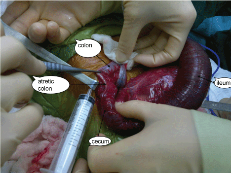
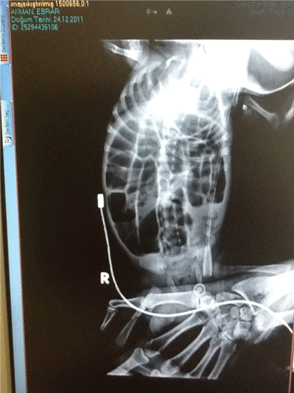
The girl baby, 28 weeks of gestation, was followed in neonatal intensive care unit because of prematurity. She has passed meconium in time. She had been feeding orally but the amount of feeding could not been increased gradually. She had developed abdominal distention on 47 days of life with sepsis findings. Sepsis criteria decreased and abdominal distention released after the sepsis treatment was begun. The baby was started to feed again, The abdominal distention has been increased over the time although the baby was very active and sepsis criteria were absent. The x-ray of abdomen (Figure: colonic atresia X-ray) showed the intestinal obstruction findings so we decided to operate on her of 69 days of life. Ascendent colon atresia was found that it was resected and made colocolic anastamosis. Pathology report proved it as a atresia. She had been discharged on the day of 18 of postoperatively. Follow-up was 2 years and she was healthy.
Colonic atresia is seen on the right side as a fibrotic cord which has a 1-2 mm lumen.
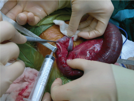
Figure 1: Colonic atresia x-ray