IMAGE ARTICLE | VOLUME 1, ISSUE 2 | OPEN ACCESS DOI: 10.23937/2474-3682/1510019
Images of Iatrogenic Pneumopericardium
Josephine Pressacco
University of Montreal, Montreal, Quebec, Canada
*Corresponding author:
Josephine Pressacco, MD, PhD, University of Montreal, 5400 Gouin Boulevard west Montreal, Quebec H4J 1C5, Canada, Tel: 514-338-2222, Fax: 514-338-354, E-mail: pressacco@gmail.com
Published: December 11, 2015
Citation: Pressacco J (2015) Images of Iatrogenic Pneumopericardium. Clin Med Img Lib 1:019. doi.org/10.23937/2474-3682/1510019
Copyright: © 2015 Pressacco J. This is an open-access content distributed under the terms of the Creative Commons Attribution License, which permits unrestricted use, distribution, and reproduction in any medium, provided the original author and source are credited.
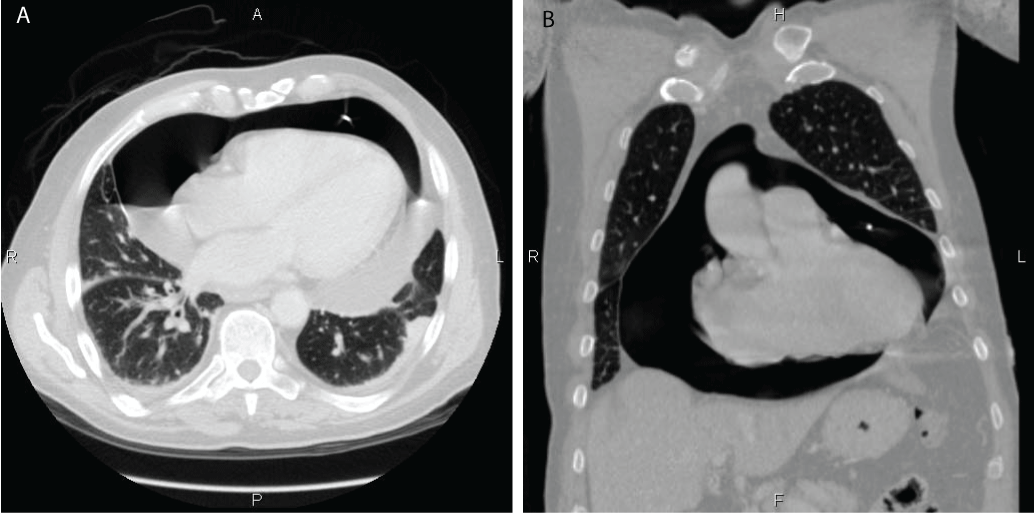
A 76-year-old man presenting with cardiac tamponade underwent urgent pericardial drainage using ultrasound guided pericardial puncture. A pericardial drainage catheter was left in place. The patient continued to present with symptoms suggestive of recurrent cardiac tamponade. Computed tomography (CT) of the chest in axial (Figure 1A) and coronal (Figure 1B) plans demonstrated a large pneumopericardium. In this particular case, the pericardial drainage catheter was malfunctioning due to a kink at its distal end. Air was entering the pericardium via the pericardial drainage catheter but could not drain out.