IMAGE ARTICLE | VOLUME 2, ISSUE 4 | OPEN ACCESS DOI: 10.23937/2474-3682/1510038
Exstrophy of Bladder
Birendra Rai1 and Prof Farhana Sharif2,3
1Pediatric Registrar, Midland Regional Hospital, Mullingar, Ireland
2Clinical Associate Professor, Royal College of Surgeons (RCSI), Ireland
3Consultant Pediatrician, Midland Regional Hospital, Mullingar, Ireland
*Corresponding author:
Birendra Rai, Pediatric Registrar, Midland Regional Hospital, Mullingar, Ireland, Tel: 3538-9411-2759, Fax: +353449394403, E-mail: drbirendrarai@gmail.com
Published: April 17, 2016
Citation: Rai B, Sharif PF (2016) Exstrophy of Bladder. Clin Med Img Lib 2:038. doi.org/10.23937/2474-3682/1510038
Copyright: © 2016 Rai B, et al. This is an open-access content distributed under the terms of the Creative Commons Attribution License, which permits unrestricted use, distribution, and reproduction in any medium, provided the original author and source are credited.
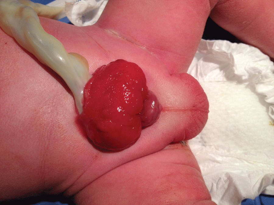
A male infant was born at 38 weeks of gestation by normal delivery. Physical examination revealed exteriorly lying bladder with deformed penis and low lying umbilicus (Figure 1). Skin over the bladder wall was deficient and mucosa of the bladder was exteriorised. A diagnosis of Exstrophy of bladder was made and urologic surgical team were contacted. He had no other associated anomaly. The swelling was dressed with saline soaked gauge and normal feeding and nursing continued. He is awaiting staged surgical closure of the defect.
Exstrophy of the bladder is seen in about 4 per 100,000 live births. It is more common in first born children with slight male preponderance. Ideally antenatal scan should detect this condition which was not detected in our case. Exteriorised posterior urethra, low lying umbilicus and anteriorly displaced anus are associated anomalies. Staged surgical repair is the treatment of choice.