Type 1 neurofibromatosis (NF-1), or Von Recklinghausen disease, is an autosomal dominant genetic disorder. It is characterized by the presence of multiple neurofibromas. Herein, we reported an interesting case of a particularly aggressive neurofibroma that resulted in lower extremity gigantism; this form of plexiform neurofibromas, called elephantiasis neuromatosa (EN) is rarely described. It usually affects the lower limbs and can be complicated by intractable pain and multiple ulcers.
Neurofibromatosis, Neurofibromas, Elephantiasis neuromatosa, Genodermatoses, Genetic skin diseases
This is the case of a twenty-eight-year-old man, with no significant family history, known to have type 1 neurofibromatosis who presented to the outpatient clinic for a growing lesion on the left lower extremity. The mass appeared during puberty and increased in size progressively during the years, affecting the quality of life of the patient. On physical exam, the lesion was soft, non-ulcerated, extending from the waist with the largest diameter reaching 30 cm (Figure 1 and Figure 2). The neuro-vascular exam was insignificant. A CT scan of the limb revealed peri-osseous dysplasia, cortical bone erosions and a low-density mass infiltrating the surrounding tissues. The MRI showed an irregular multilobed infiltrating lesion with increased intensity on T2W/STIR and targetoid images, suggesting the diagnosis of a plexiform neurofibroma and elephantiasis neuromatosa. Additionally, the patient had 5 Café-au-lait macules (> 15 mm of diameter), multiple neurofibromas located on the trunk and extremities (Figure 2), bilateral axillary freckling. The ophthalmologic exam showed hamartomas of the iris. A surgical consult was ordered. The patient was subsequently lost to follow-up.
Type 1 neurofibromatosis (NF-1), or Von Recklinghausen disease, is an autosomal dominant genetic disease that affects 1 in every 3000 newborn [1]. Besides cafe-au-lait macules, axillary and inguinal freckling and bone/ophtalmological abnormalities, neurofibromas are considered one of the key findings in this disease [1]. Plexiform neurofibromas are indeed diagnostic of NF-1 and represent a major cause of morbidity and mortality. They are found in 30-50% of NF-1 patients, appear in childhood and carry a risk of malignancy estimated around 5-10% [2,3]. Herein, we reported an interesting case of a particularly aggressive neurofibroma that resulted in lower extremity gigantism; this form of plexiform neurofibromas, called elephantiasis neuromatosa (EN) is rarely described. It usually affects the lower limbs and can be complicated by intractable pain and multiple ulcers [4]. MRI findings include the typical targetoid images seen on T2W/STIR sequence [4]. Pathological findings include thickened nerve fascicules with myxoid stroma and fibroblast cords interlaced with Schwann cells [3]. The mainstay of treatment is surgery with high risk of bleeding and recurrence [4]. It is indicated in case of intractable pain, neurological or vascular deficiency, or malignancy [4]. Treatment with Imatinib, Interféron-alpha or Rapamycin is still controversial [5].
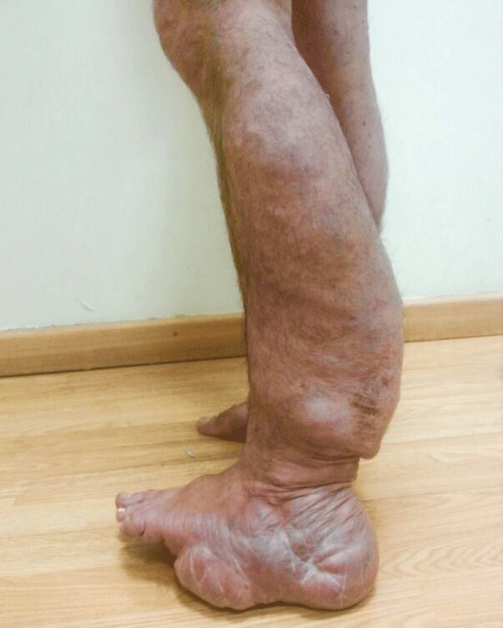
Figure 1: Left lower extremity elephantiasis neuromatosa.

Figure 2: Multiple neurofibromas on the trunk and extremities.