Intestinal metastasis are a common finding in patients affected with melanoma; only exceptionally they can cause enteric intussusceptions and present clinically as bowel obstruction. We present the case of a young woman admitted to our institution for an acute bowel obstruction caused by multiple intestinal invaginations from melanoma metastasis.
Melanoma, Ileal melanoma, Ileal intussusceptions, Intestinal occlusion
We present the case of a woman of 38 years with no relevant medical history, except for the removal of two benign epidermal naevi five years earlier, who was admitted to our surgical ward with abdominal pain and vomiting. She underwent abdominal X-Ray and CT-Scan that identified a suspect ileal volvolus. We executed a diagnostic laparoscopy and found one ileocolical invagination that required conversion to laparotomy for the management. We identified multiple blackish nodular lesions of 1-3 cm of size emerging from the sierosa through all the length of the jejunum and ileum determining four additional small bowel invaginations that were reduced manually (Figure 1). The ileo-ciecal intussusception was instead resected because of signs of ischemia; at the opening of the specimen the serosal nodular lesion was found to have also an intramucosal infiltration (Figure 2). The histological diagnosis was ileal melanoma with BRAF activating mutation and metastasis to the mesenteric lynphnodes. The subsequent stadiation through FDG-PET-CT scan revealed multiple metastases in the chest, abdomen, mediastinum and bones; no cutaneous or uveal primitive lesions were found. Postoperative course was uneventful and the patient was referred to oncological treatment with BRAF inhibitors. Intestinal metastasis are a common finding in asymptomatic melanoma patients; only exceptionally they can cause enteric invagination and present clinically as an acute bowel obstruction [1-4].
All authors equally contributed to this paper with conception and design of the study, literature review and analysis, drafting and critical revision and editing, and final approval of the final version.
No potential conflicts of interest. No financial support.
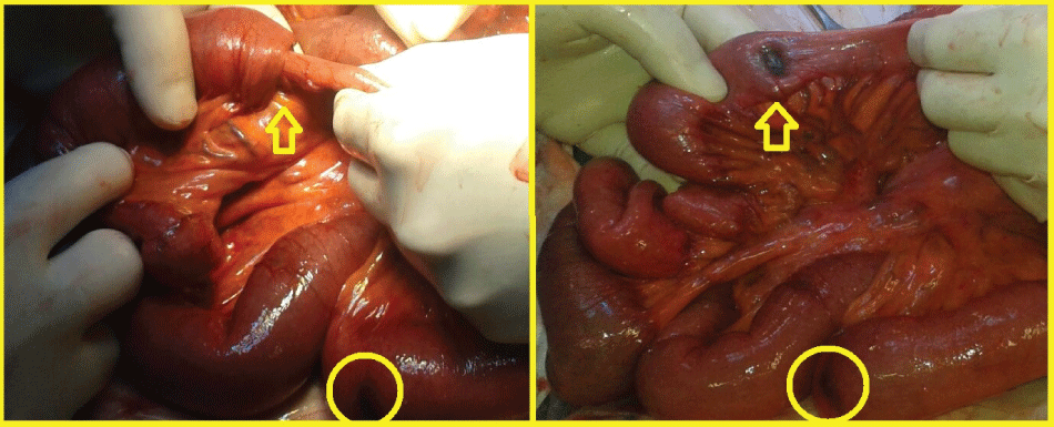
Figure 1: One small bowel invaginations that was reduced manually. A blackish node was found in intussusception.
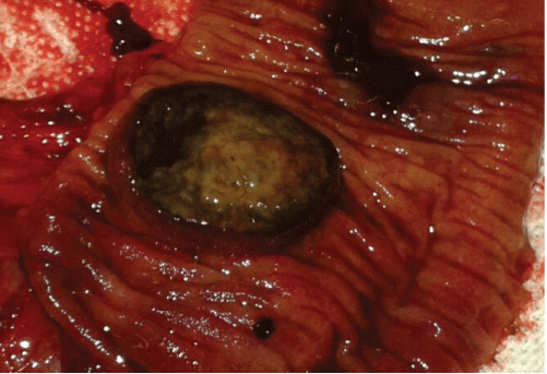
Figure 2: Ileo-ciecal intussusception that was resected. Nodular lesion was found to have also an intramucosal infiltration.