A 66-year-old male patient who underwent operation on the left temporal region due to squamous cell cancer and has been followed for a year admitted to our hospital for right ear swelling and ear pain which is worse at night. Physical examination showed patient had slight swelling of the right auricular helix. Laboratory findings was normal. Contrast magnetic resonance imaging (MRI) is performed on patient with history of malignancy because of considering metastasis. MRI showed the lesion size was 15 × 10 milimeters, in the region corresponding to right auricular helix. It was hypointense on T1-weighted images and hyperintense on T2-weighted images. After ıntravenous injection of contrast agent; lesion was enhanced and demonstrated diffuse increase in thickness of right auricula (Figure 1). Described lesion has been surgically removed from the patient who had primary malignancy background. Histopathological assessment of the lesion demonstrated epidermal acanthosis, granulomatous dermal inflammation and dermal thinning; hence the diagnosis of chondrodermatitis nodularis chronica helicis is confirmed [1-4].
Chondrodermatitis nodularis chronica helicis (CNCH) is a benign inflammatory disorder, it is characterised with auriculer nodul. CNCH often occured on the auricular helix, less frequently on the auricular antihelix, it is characterised with painful nodul or nodules. CNCH is first described by Winkler, in 1915; for this reason also known as Winkler's disease. Although the pathological diagnosis is easy with it's specific histopathological findings, this pathology is less known by radiologists. In such cases, similar to our patient, the disease may be confused with metastasis especially in the differential diagnosis of patients with primary malignancy that arises from the skin. We described magnetic resonance imaging (MRI) findings of a 66-years-old man with painful tumor at helicis of right ear who have history of squamous cell cancer on the left temporal region skin [5,6].
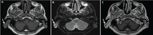
Figure 1: Axial T1 weighted MRI of the right auricular helix (A) demonstrates hypointense nodular mass lesion. On axial T2 weighted MRI; (B) it appears as hyperintense nodular mass lesion. Contrast enhancement is not appreciated at contrast-enhanced T1-weighted imaging; (C) diffuse increase in thickness of right auricula was visible on all sequences.