The introduction of an exogenous pigment in the dermis is not without risk. We report a case of a sarcoidosis complicating a traditional tattoo after several years. Currently only temporary henna tattoos are subject to frequent publications in the literature. It must be known to the dermatologist ahead this type of skin lesions will have to undertake explorations necessary for the identification of systemic sarcoidosis.
Traditional tattoo, Sarcoid nodules
The traditional tattoo in Morocco is considered one of the oldest rituals of the berber culture. The tendency of sarcoid granulomas to infiltrate old scars and tattoos is well documented. It represents one of "allergic" reactions to ink or colouring agents, which constitute the main current complication associated with tattoos that lead individuals to consult. We report, to our knowledge, the first case of sarcoidosis on a traditional tattoo.
A 57-year-old woman, presented with papular erythematous-purplish lesions, indurated and asymptomatic, evolving for one year, along traditional permanent tattoos done for over 30 years (Figure 1), of the chin and wrist of the right hand. Physical examination was normal, no respiratory symptoms were found. The lesions were suggestive of sarcoidosis, a foreign body granuloma, or pseudolymphoma. Dermoscopical examination showed translucent yellow to orange globular like, with linear vessels; these lesions were suspecting the presence of granuloma (Figure 2). Histological examination showed non caseating granulomas. The diagnosis of sarcoidosis was made on clinical, dermoscopical and histological arguments. The systematic assessment was normal. A treatment with topical corticosteroid was introduced for a month with good improvement.
Sarcoidosis is a systemic granulomatous disease of undetermined cause. The sarcoid granulomas have often a tropism for old scars and tattoos, foreign material inoculation sites [1]. Several cases of sarcoidosis have been described on tattoo [2], but no cases of sarcoidosis on a traditional tattoo have been reported up to now in the literature. Complications tattoos concern from 2% to over 20% of subjects tattooed. It is important, especially in our context, where these tattoos are common and usually hidden, to detect signs of sarcoid granulomatous complications especially to diagnose and search for systematic signs at an early stage.
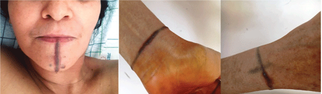
Figure 1: Papular erythematous-purplish lesions of the chin and wrist of the right hand.
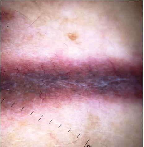
Figure 2 : Dermoscopical examination: translucent yellow to orange globular like, with linear vessels.