Kallmann syndrome, Olfactory, MRI
A 14-years-old male was consulted for cryptorchidism and anosmia. There was decrease of LH, FSH and testosterone in the laboratory examinations of the patient. We used Magnetic Resonance Imaging (MRI) to visualize the olfactory tract and evaluate the olfactory sulci in patient whose clinical and laboratory findings were compatible with Kallmann syndrome. Coronal images of the frontal region clearly demonstrated aplasia of the bilateral olfactory sulci and absence of the olfactory tracts in the patient (Figure 1).
Kallmann syndrome is a neuronal migration disorder characterised by hypogonadotropic hypogonadism and anosmia or hyposmia [1]. It is generally accepted that defective rhinocephalon development result in olfactory tract abnormalities [2]. Cases generally are admitted with hyposmia, anosmia and puberte tarda [3,4]. MRI is very successful that show the hypoplastic or aplastic olfactory sulci and absence olfactory bulbs and tracts. A hypoplastic anterior pituitary may also be seen. There are other congenital disorders associated with decreased olfaction, such as holoprosencephaly, Down syndrome and Turner syndrome. But complete absence of the olfactory bulbs and tracts has been defined only in Kallmann syndrome [3]. In conclusion, MRI is a useful method to demonstrate abnormalities of the olfactory system which are always present among patients suffering from Kallmann syndrome.
None.
None.
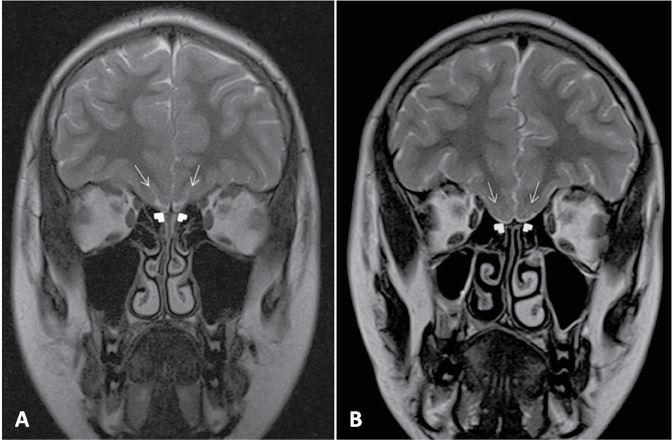
Figure 1: Coronal T2 weighted images. A) Normal MRI finding of bilateral olfactory sulcus (arrows) and olfactory bulbs (arrowheads); B) In the patient with Kallmann syndrome, bilateral olfactory sulcus (arrows) and olfactory bulbus cannot be seen (arrow heads).