Diastematomyelia, Spinal cord, Vertebrae, Magnetic resonance imaging
A 7-year-old male was brought with a marked spinal deformity. Physical examination showed rotational scoliosis and loss of lumbar lordosis. The patient was referred for a Magnetic Resonance Imaging (MRI) of the lumbosacral spine. MRI showed a left-convex rotational scoliosis and loss of lumbar lordosis. There was a fusion of T10-T11-T12 vertebral bodies with a partial excision (Figure 1). Spinal canal was widening at the level L2-L4. The terminal cone was reaching L5 level. At the level of T10 spine, the spinal cord was divided into two hemicords and two dural tubes up to the upper part of the L3 body. There was a bone septum, dividing the spinal canal into two separate canals including two hemicords in separate dural tubes (Figure 2).
Diastematomyelia is a rare congenital abnormality of the spinal cord in which a part of the spinal cord is split in the sagittal plane into two hemicords [1]. It is more common in women (between 80 to 90%) and usually appears within the thoracolumbar spine (85%) [2]. Two hemicords, each covered by an intact layer of pia arachnoid, travel through a single subarachnoid space surrounded by a single dural sac in nearly 60% of patients with diastematomyelia [1,2]. Among 40% of patients, a bony spur or a fibrous band passing through the two hemicords is encountered [2]. This rare congenital malformation of the spinal cord, which belongs to the group of occult spinal dysraphisms. It may coexist with other spinal dysraphisms, such as myelomeningocele, meningocele, spinal lipoma, neuroenteric cysts or dermal sinuses and vertebral abnormalities, such as hemivertebrae, butterfly vertebrae or scoliosis [3]. Prior to the universal availability of MRI scan, myelography and CT were used to add a new dimension to the diagnosis of dysraphic lesions of the spine [1,3].
None.
None.
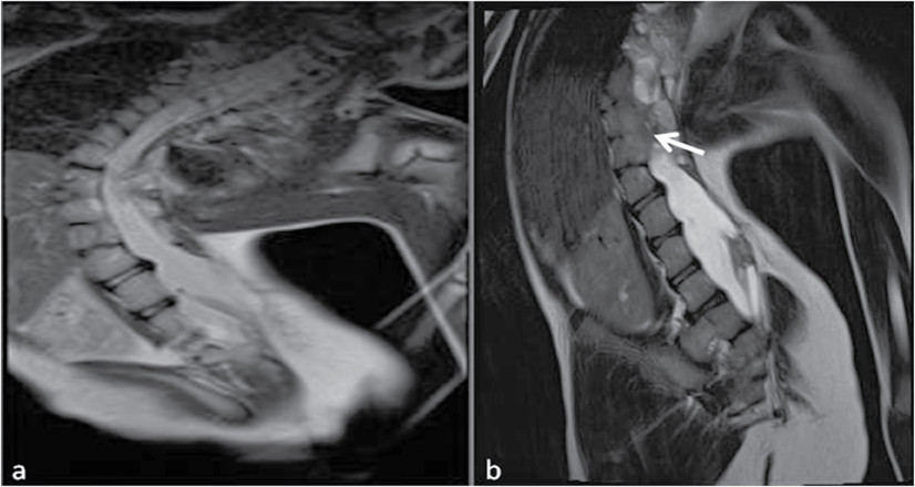
Figure 1: Coronal T2-weighted (a) and sagittal T2-weighted (b) MR images show left-convex rotational scoliosis and fusion of T10-T11-T12 vertebral bodies with a partial excision (arrow).
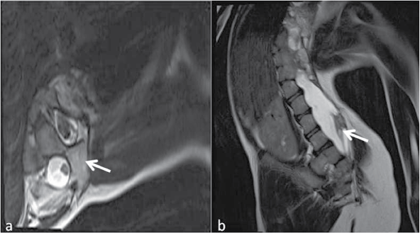
Figure 2: Axial T2-weighted (a) and sagittal T2-weighted (b) MR images show a bone septum, dividing the spinal canal into two separate canals including two hemicords in separate dural tubes and the terminal cone reaches the L5 level (arrows).