IgG4-related disease is a relatively new disease which has gained significant recognition in the last decade. It is an immune mediated disease and diagnosis is often missed because of limited awareness and a typical presentation. The diagnosis of IgG4-related disease is confirmed by a characteristic histopathological appearance in any organ site, comprising of lymphoplasmacytic infiltrate, storiform fibrosis and obliterative phlebitis with elevated IgG4-positive plasma cells. Early detection is crucial to prevent organ damage as they are usually responsive to glucocorticoids and immunomodulatory therapy.
IgG4 related disease, Lymphoplasmacytic infiltration, Storiform fibrosis, IgG4, Glucocorticoids
A 50-year-old lady presented with a one month history of progressively enlarging, painful left orbital swelling. She had failed outpatient oral antibiotic therapy. Physical examination was significant for left periorbital and orbital swelling, proptosis, mechanical ptosis, tenderness, edema, erythema and warmth. Visual acuity was 20/20 with restriction of extraocular movements. Laboratory workup showed normal white cell count, erythrocyte sedimentation rate, elevated C reactive protein and positive wound culture growing rare leukocytes and Staphylococcus aureus. Magnetic resonance imaging of the orbits and face showed left orbital cellulitis with subperiosteal enhancement, erosive changes in the ethmoid air cells and lateral displacement of left globe (Figure 1). She was started on broad spectrum antibiotics on which she failed to improve. Diagnostic left caruncular orbitectomy with biopsy was done which showed dense lymphoplasmacytic infiltrate, fibrosis and many eosinophils (Figure 2 and Figure 3). Immunohistochemical studies revealed diffusely positive > 100 IgG4 cells/HPF (Figure 4). This was diagnostic for IgG4 related disease [1]. Serum IgG4 level was only mildly elevated at 129 mg/dL. She was started on intravenous methylprednisolone 60 mg with dramatic improvement in symptoms and was discharged on prednisone.
IgG4-related disease is a relatively new systemic immune mediated disease which affects multiple organs [2]. It has gained significant recognition in the last decade but diagnosis is often missed because of limited awareness and atypical presentation. Hallmark histopathological findings include lymphoplasmacytic infiltration, storiform fibrosis and obliterative phlebitis with modest eosinophilia [3]. These diseases are generally responsive to glucocorticoids and early recognition is crucial to prevent organ damage [4]. In cases refractory to steroids, rituximab is used [5].
We thank Dr Alexandra Buddhai for providing us with the histopathology images.
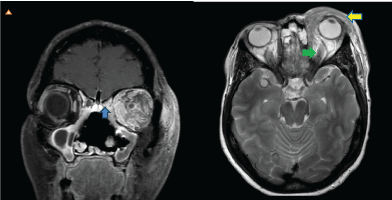
Figure 1: MRI coronal T1 post contrast with fat saturation showing left sided subperiosteal enhancement and erosion of the adjacent ethmoid air cells [blue arrow]. Axial enhanced fat suppressed T2 MRI showing preseptal [yellow arrow] and postseptal enhancing tissue with lateral displacement of globe [green arrow].
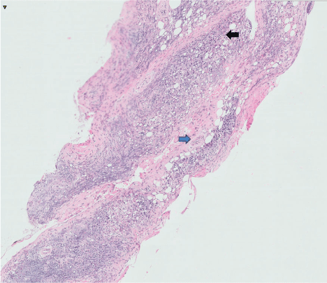
Figure 2: Histology showing lymphoplasmacytic infiltrate [black arrow] and fibrosis [blue arrow].
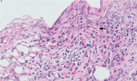
Figure 3: Histology revealing eosinophilia [blue arrow] and plasmacytoid lymphocytes [black arrow].
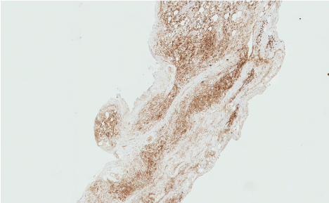
Figure 4: Histology showing diffuse staining with IgG4 positive cells [black arrow].