Inguinal mass, Testicular mixed germ cell tumor, US, MRI
A 19-year-old man was admitted our hospital with right inguinal mass. Ultrasonography (US) showed an irregular, heterogeneous mass lesion in the right ingunal region. Right testis was not seen in scrotum. The patient had no history of any operation. Magnetic resonance imaging (MRI) demonstrated a giant mass which was size of 24 × 17 × 15 centimeter. The mass was hypointense on T1-weighted images, heterogeneous hyperintense on T2-weighted images and heterogeneous contrast enhancement was observed (Figure 1). The lesion was invading the surrounding fat and muscle tissues and also prolonged in the intraabdominal space. The absence of the right testis suggested a testis tumor. Soft tissue sarcoma was considered in differential diagnosis. The lesion was excised and resulted as testicular mixed germ cell tumor (GCT) histopathologically.
GCT of the testes are the most common malignant tumor in males who are between 15 and 44-years-old. GCTs are classified as either nonseminomatous which account for approximately 60% of testis tumors, or seminomas which account for approximately 40% [1]. Nonseminomas are clinically more aggressive and often include multiple cell types [2,3]. Risk factors for the development of testicular GCT include cryptorchidism, infertility, testicular dysgenesis and a positive family history [1,4]. In conclusion, when an inguinal mass is detected in a young adult male patient, the story of cryptorchidism should be questioned, scrotal US should be performed and should be kept in mind that it may be originated from testis.
None.
None.
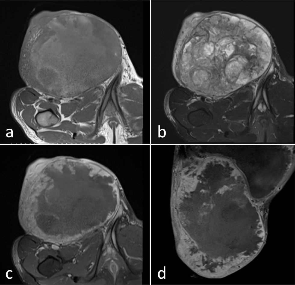
Figure 1: MRI shows an irregular, heterogeneous mass lesion in the right ingunal region. The mass is hypointense on T1-weighted images (a), heterogeneous hyperintense on T2-weighted images (b) and after contrast adminstration (c,d) heterogeneous contrast enhancement is observed.