Aneurysmal bone cyst, Sacrum, MRI
A 20-year-old man presented with a 2-year history of severe pain in his sacral region. He described an intermittent pain of moderate to severe intensity, localized in the left flank region, increasing at night. Pain is partially relieved by rest, supine position and is lately associated with a limp. Magnetic Resonance Imaging (MRI) revealed an expansive destructive cystic lesion at the S1, S2 and S3 vertebral levels, lesion's close association with S1 and S2 sacral nerve roots, and extending posteriorly into the spinal cord leading to the narrowing of the spinal canal. The lesion appeared hypointense on T1-weighted images and hyperintense on T2-weighted images. The fluid-fluid level was not observed inside the lesion, but limited areas have T1 hypointense T2 hyperintense blood elements (Figure 1). Biopsy established the diagnosis of Aneurysmal Bone Cyst (ABC).
ABC is a destructive, expansile, locally agressive tumor-like lesion of the bone characterized by a reactive proliferation of connective tissue containing blood filled cavities [1]. ABC is representing 1% of all primary benign bone tumors, with low risk of malignant transformation [2,3]. ABC most commonly occur in the distal femur or proximal tibia. The pelvis and posterior elements of the spine are also commonly involved. Sacral location is very rare [3,4]. In sacral location, therapy is limited by lesion's close association with sacral nerve roots and the possibility of resultant neurologic deficit [3]. A differential diagnosis from chordoma, osteoblastoma and giant cell tumor is required [2-4]. MRI is the optimal modality for evaluating the contents of the ABC as well as the full extent of the bone and soft tissue involvement, including the degree of compression of the neural elements [4]. The presence of fluid-fluid interfaces on the T2 weighted images is characteristic and highly suggestive of an ABC but this sign is not specific.
None.
None.
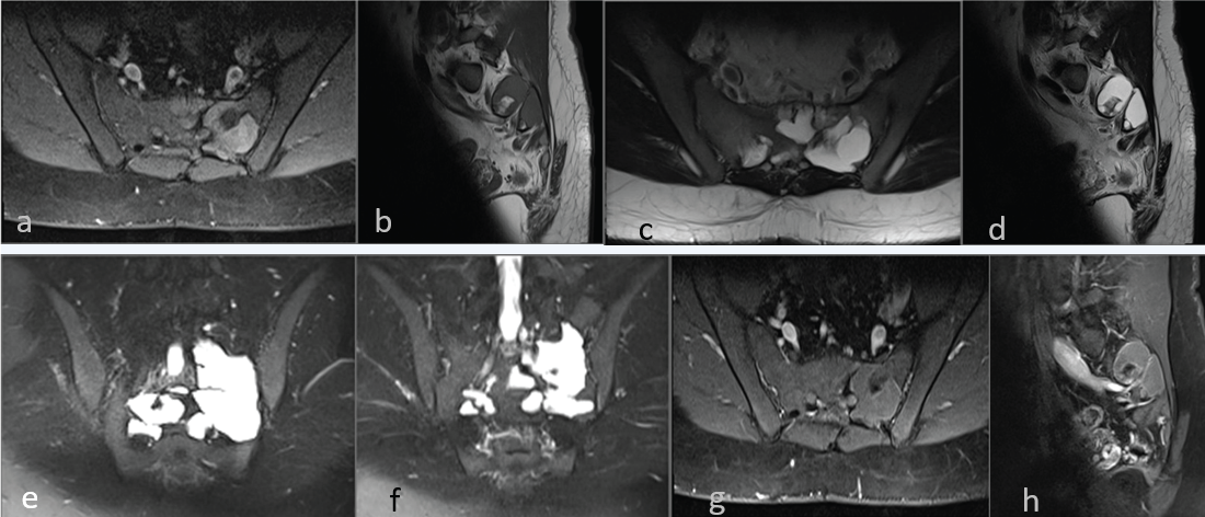
Figure 1: a) T1-w axial fat-sat; b) T1-w sagital; c) T2-w axial; d) T2-w sagittal; e) T2-w fat-sat coronal; f) T2-w fat-sat coronal; g) T1-w fat-sat axial; h) T1-w fat-sat sagital. MR images of patient. There is an expansive destructive cystic lesion at the S1, S2 and S3 vertebral levels, lesion's close association with S1 and S2 sacral nerve roots, and extending posteriorly into the spinal cord leading to the narrowing of the spinal canal. The lesion appeare hypointense on T1-weighted images and hyperintense on T2-weighted images. There are blood elements, limited areas inside the lesion (T1-w hypointense T2-w hyperintense).