Deep Vein Thrombosis (DVT) occurs when there are clots in the deep veins of the body, accounting for the majority of venous thromboembolism (VTE) cases. Half of the cases may report symptoms of pain, swelling, warmth and discoloration or redness which may be similar to cellulitis. Here we report a 47-year-old Filipino female who presented with bullae on her left leg, associated with pain, swelling and erythema which improved with anticoagulants and antibiotics.
Deep vein thrombosis, Cellulitis, Filipino
Deep vein thrombosis (DVT) is an important and common disease, being the most common cause of death from cardiovascular diseases, it is reported to occur in 100 per 100,000 in the general population [1]. Lower extremity DVT usually begins in the calf and propagates proximally to the popliteal vein, femoral vein, and iliac veins. In about half of the cases, there will be swelling, warmth and discoloration or redness which may be similar to cellulitis. Cellulitis is an infection of the deep dermis and subcutaneous tissue, most often caused by bacteria, that presents with the classic signs of inflammation [2]. The occurrence of both deep vein thrombosis with cellulitis is rare, hence in this case report, we will highlight the manifestations of both conditions found in our patient. Limited cases with an overlap of DVT and cellulitis have been reported, and no cases from the Philippines had been published in literature yet.
We report a 47-year-old Filipino female, who months prior, had been apparently well, without known comorbidities until 1 month when she noted occasional numbness of the left heel. She then noted swelling of her left knee with a pain scale of 4-5/10, associated with the onset of gradual swelling, erythema and warmth on her left leg, more prominent on the posterior aspect, up to the dorsum of the foot. Arterial and venous duplex scan was done which revealed thrombosis at the left peroneal vein; the Ankle-Brachial Index was also noted to be > 2.5 (noncompressible arteries) [3] for both lower extremities. The patient was then admitted for further management.
Three days after, vesicles erupted, evolving into bullae on the same sites, rupturing and leaving desquamation of the superficial skin, with crusting and purulent discharges (Figure 1). Blood tests showed leukocytosis with neutrophilic predominance, and elevated C-reactive protein levels. Using the ALT-70 Model for Cellulitis, the patient had a score of 5 which had a 95% likelihood of cellulitis [4]. Wound gram stain and culture sensitivity test noted growth of Staphylococcus aureus . This supports the diagnosis of the patient having concomitant cellulitis on top of DVT. The patient was then managed with intravenous antibiotics, dressings with NSS compress, silver sulfadiazine and gentle debridement of the crusts with a once-a-day intravenous dose of enoxaparin. With improvement, the enoxaparin was shifted to oral Apixaban. The patient was then discharged with home medications, with lesions fully resolving after 1 month (Figure 2).
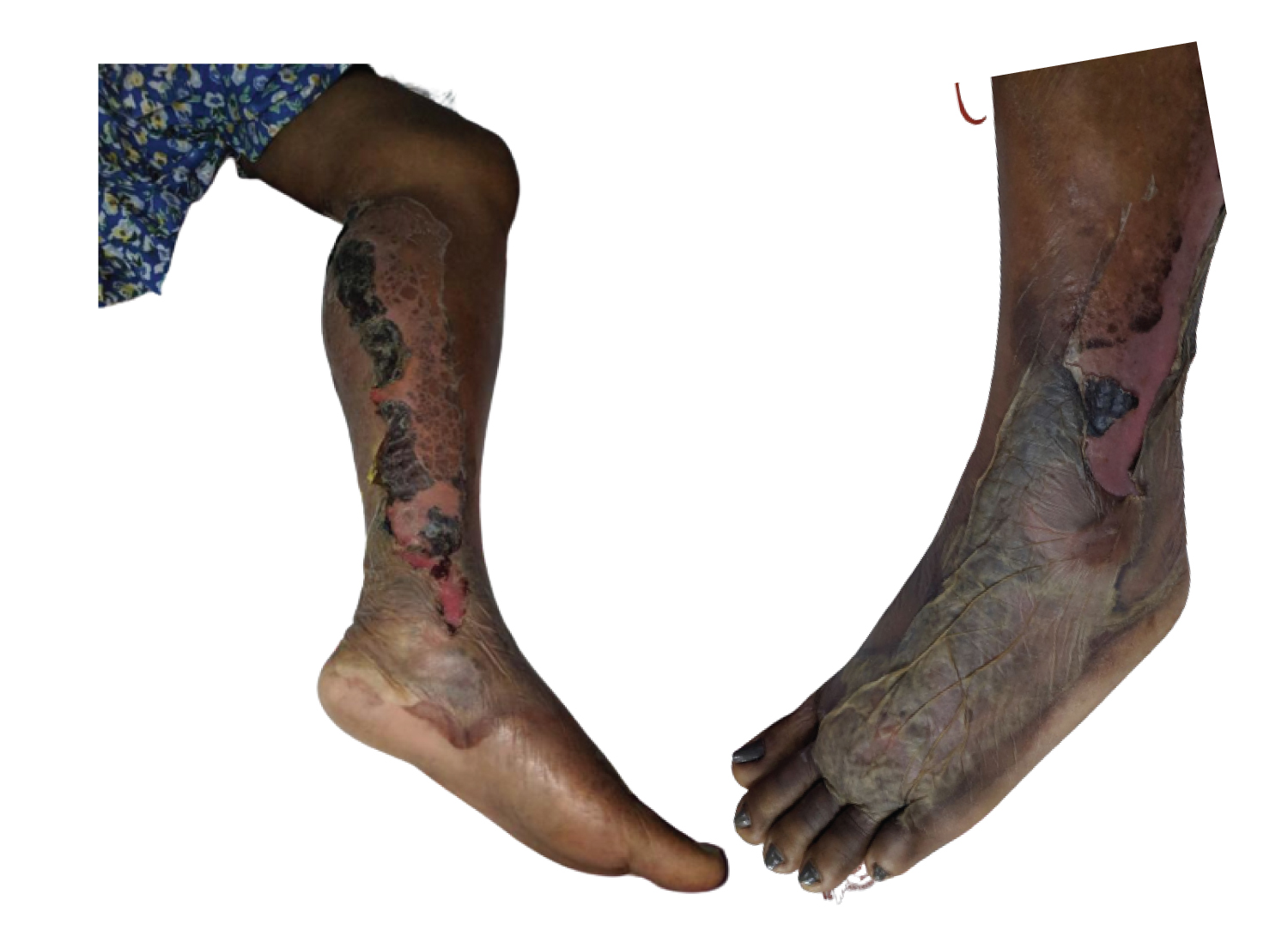 Figure 1: Bullae formation on the medial aspect of the left leg, extending to the dorsum of the left foot.
View Figure 1
Figure 1: Bullae formation on the medial aspect of the left leg, extending to the dorsum of the left foot.
View Figure 1
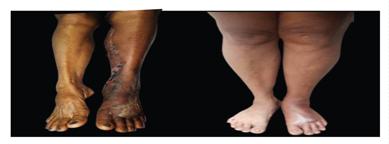 Figure 2: Before and after 1 month of treatment.
View Figure 2
Figure 2: Before and after 1 month of treatment.
View Figure 2
Venous thromboembolism (VTE) is a condition encompassing deep venous thrombosis (DVT) and pulmonary embolism. The annual incidence of VTE is estimated to be 1-2 individuals per 1000, with approximately two-thirds of cases attributed to DVT alone [5,6]. While incidence rates in Asian populations were historically lower than in Western countries, they have been increasing over time [7].
Thrombosis, a protective mechanism that prevents blood loss, can become pathological and lead to thrombus propagation, causing vessel blockage. Virchow identified three mechanisms involved in DVT: Damage to the vessel wall, blood flow turbulence, and hypercoagulability. The most common sites of DVT are the lower limbs, particularly below the knee, including low-flow areas such as the soleal sinuses and venous valve pockets. Various risk factors contribute to DVT, including prolonged inactivity, cancer, surgery, inherited coagulation disorders, heart failure, pregnancy, obesity, smoking, and the use of oral contraceptives [4].
Symptoms of DVT include edema, erythema or hyperpigmentation of the skin, pain, and occasionally ulcers [1,4]. However, the development of vesicles and bullae on the affected area is a rare and acute manifestation resulting from acute changes in interstitial pressure due to vein blockage. This leads to the separation of epidermal cells at the level of the stratum spinosum, causing extensive bullae due to fluid retention [4,8].
The National Institute for Health and Care Excellence (NICE) guidelines recommend treating a positive proximal leg vein ultrasound as a case of DVT [4]. The first-line treatment for DVT and other VTEs includes anticoagulants such as apixaban or rivaroxaban, which are highly selective direct factor Xa inhibitors, acting on both free and clot-bound factor Xa [9].
Cellulitis, an inflammatory condition resulting from dermis and hypodermis infection, shares several clinical characteristics with DVT. However, there are differences in their clinical presentation. Cellulitis typically presents with ill-defined borders, a dusky hue, surface breaks with vesicles, and may progress to hemorrhagic bullae, necrotic tissue discharge, or adherent crusts [10,11]. The diagnosis of cellulitis relies mainly on clinical history and physical examination, as there are no reliable diagnostic studies. The ALT-70 Cellulitis Predictive Model can aid in differentiating cellulitis from other similar conditions. In contrast, pustules, abscesses, or fluid collections associated with purulent cellulitis should be drained and cultured early during diagnostic workup [2].
In this case, although the patient already presented and was diagnosed with DVT, the clinical presentation warranted further investigations. The occurrence of DVT with cellulitis is rare, and only documented in a few case reports, aside from the fact that cellulitis is also a differential diagnosis for DVT. Patients with DVT and cellulitis may exhibit similar symptoms such as erythema, edema, tenderness of the affected limb, and low-grade fever. However, raised blood counts, inflammatory markers, and blood cultures can aid in differentiating between the two conditions. This further establishes that both conditions were found in the patient as evidenced by the thrombosis of the left peroneal vein on Doppler ultrasound, and the positive culture result from the wound drainage of the same leg.
The presence of conditions that present similar symptoms, such as deep vein thrombosis (DVT) and cellulitis [12], serves as a reminder for medical professionals to prioritize the use of appropriate diagnostic and treatment strategies. When patients exhibit symptoms of a hypercoagulable state, such as leg pain, erythema, and edema, which could potentially be indicative of cellulitis or co-existent with it, it becomes crucial to conduct further investigations. By conducting thorough examinations and considering additional factors, healthcare providers can ensure accurate diagnosis and implement suitable treatment approaches for the patient's condition.
No funding for this case report.
Both authors have no conflict of interest.