The vestibular papillae (VP) represent an anatomical variant of the normal genital epithelium and are probably the female equivalent of pearly penile papules. It is thought that they are present in 1% of women, and this low percentage may be due to their lack of knowledge by practitioners since their diagnosis are often wrongly spotted as condyloma acuminates warts and this can lead to aggressive investigations, therapy, and anxiety in patients hence the role of dermoscopy. Here, we present a case with 39 weeks of pregnancy, who is planned to undergo cesarean section due to a wrong diagnosis of genital warts. Dermoscopy was performed to differentiate VP from condyloma and the patient gave birth vaginally without any problem.
A correct diagnosis of vestibular papillomatosis prevents aggressive investigations and unnecessary therapies. Therefore, it is worthwhile to draw the attention of dermatologists to this entity.
Vestibular papillomatosis, Dermocopy, Condyloma
Vulvar vestibular papillomatosis (VVP) is a benign anatomical variant of the vulva [1]. The prevalence is 1-33% with the highest between 21 and 56 years at reproductive ages [2,3]. The first description of this condition was in 1981 by Altmeyer [1,2]. Since then, they have been reported under a variety of names: Hirsutoid papillomas of vulva, vulvar squamous papillomatosis, micropapillomatosis labialis, and squamous vestibular micropapilloma [4]. The lesions most often are asymptomatic, though some women describe an associated itching or burning sen-sation [5].
In pregnant women, misdiagnosis of these harmless lesions as genital warts may lead to cesarian section, because of the risk of laringeal papillomatosis in infants born vaginally to mothers with active HPV infection. This condition can cause complications associated with invasive interventions and delay in the connection of the mother with the newborn.
Twenty years woman old with 39 weeks of primigravid pregnancy was referred by a gynecologist for evaluation of suspected genital warts. The medical history of the case showed that swelling was developed at 35th weeks of pregnancy without symptoms. The patient had no suspicious sexual contact. Physical examination of the area showed multiple soft filiform papules, which are symmetrically located at the inner side of labia minor with a same color with mucosa (Figure 1). Acetowhitening was not detected after application of 5% acetic acid. In front of this aspect, diagnosis of vulvar physiological papillomatosis was suspected. In dermoscopy, these projections were pink like the rest of the mucosa, regular, symmetrical and linear, juxtaposed and their respective bases remained separate white arrows. Abundant and irregular linear vessels were observed in the transparent nuclei of the papillae (Figure 2). Patient did not give permission to take a biopsy from the lesion. Therefore, the lesion was considered as VP after evaluating the findings obtained from the history, examination, acetic acid application and dermoscopy. Spontaneous vaginal delivery was occurred at 41th weeks of pregnancy without complications.
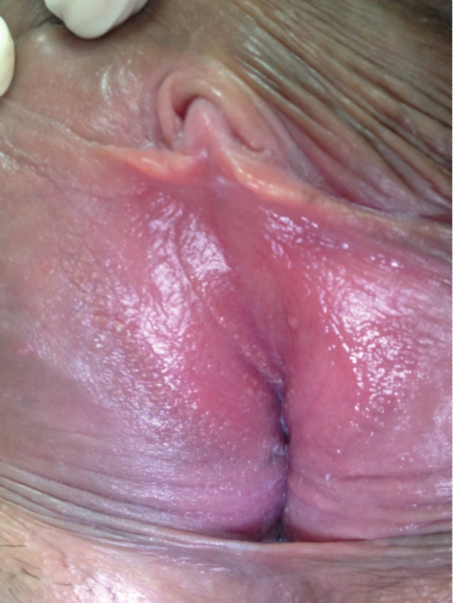 Figure 1: Clinical image showing: Soft, filiform projections with symmetrical arrangement along both sides of the vulvar vestibule.
View Figure 1
Figure 1: Clinical image showing: Soft, filiform projections with symmetrical arrangement along both sides of the vulvar vestibule.
View Figure 1
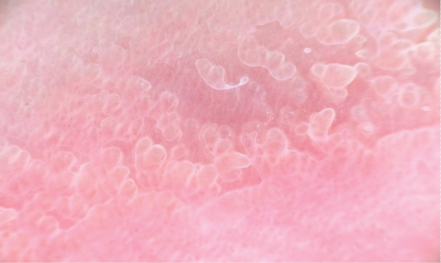 Figure 2: Dermoscopycal image showing pink, regular and linear projections, juxtaposed with separate bases. Abundant and irregular linear vessels in the transparent nuclei of the papillae.
View Figure 2
Figure 2: Dermoscopycal image showing pink, regular and linear projections, juxtaposed with separate bases. Abundant and irregular linear vessels in the transparent nuclei of the papillae.
View Figure 2
The clinical resemblance and localization of vestibular papillae has caused controversy about its etiology. Vestibular papillae have been reported with HPV infection, but a consistent association has not been proven. Moyal-Barranco and al have refuted this theory and reported that vestibular papillae were not related to HPV infection [5]. Using molecular hybridization, they detected HPV DNA sequences in only two (6.9%) of the 29 specimens of vestibular papillae, compared to 96% of specimens from vulvar warts [5]. Most recent studies have confirmed this result and shown that HPV infection is not a cause of vestibular papillomatosis [6]. Five clinical parameters were suggested by Moyal-Barranco in order to facilitate the differential diagnosis of VP from genital warts (Table 1) [5]. Vestibular papillae are symmetric or linear, soft, and pink-colored. The bases of individual vestibular papillae projections remain separate. On the other hand, condyloma acuminatum is hard and irregular. Individual projections can coalesce in a common base. In addition, in most cases, condyloma acuminatum exhibits whitening when subjected to the acetic acid test [5].
Table 1: Clinical differential diagnosis with vestibular papillae and condyloma accuminata. View Table 1
Dermatoscopy of vestibular papillomata has been defined by Su-Han Kim, et al. [7]. It reveals abundant and irregular vascular channels in the transparent core of uniform-sized cylindrical papillae. These papillae are pink like the rest of the mucosa, regular, symmetrical and linear juxtaposed with separate bases (Figure 2). However, dermatoscopy of condyloma acuminatum (Figure 3) shows multiple, irregular projections with tapering ends arising from a common base. The projections have glomerular conglomerate vascular structures with hemorrhages (small red or black dots or striae), and are more white and broader than vestibular papillae; this might correlate with the hyperkeratotic and acanthotic features of condyloma acuminate [7,8]. Therefore, along with the five clinical parameters suggested by Moyal-Barranco, a characteristic dermatoscopic finding provides additional diagnostic clues to differentiate vestibular papillae from condyloma acuminate [7].
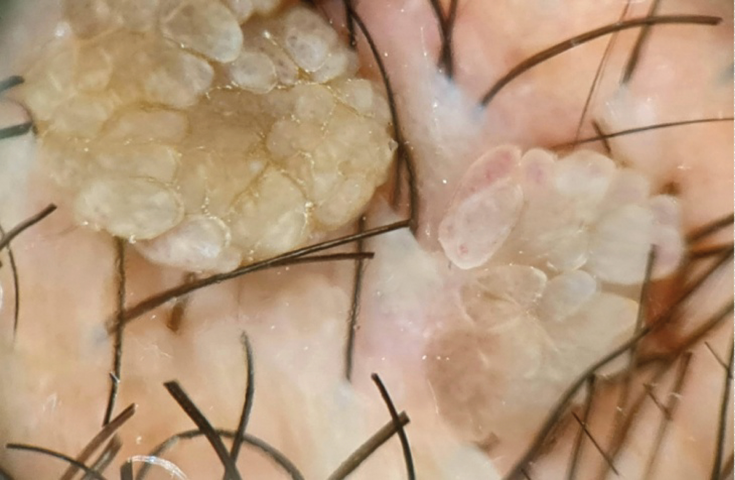 Figure 3: Dermoscopycal image of condyloma acuminate showing multiple irregular projections with tapered ends arising from a common base, whiter and wider than the vestibular papillae with conglomerate vascular structures.
View Figure 3
Figure 3: Dermoscopycal image of condyloma acuminate showing multiple irregular projections with tapered ends arising from a common base, whiter and wider than the vestibular papillae with conglomerate vascular structures.
View Figure 3
Because vestibular papillae are unfamiliar to clinicians, they may be misdiagnosed as condyloma acuminata, leading to inappropriate treatment. Dermatoscopy may represent a convenient and helpful modality in the diagnosis of vestibular papillae, especially in a pregnant woman.