Extramedullary presentation of acute undifferentiated leukemia is uncommon, and it is unclear if this is an initial manifestation of a subsequent systemic disease. We present a 67-year-old Caucasian female with skin lesions and multiple enlarged lymph nodes. Microscopy showed diffuse leukemic infiltrates, positive for dim CD45, CD34, CD117, TdT, CD56 and CD43, but negative for HLADR, MPO, CD4, CD68, CD3, CD4, CD20, CD19, and CD79a. We discuss the differential diagnosis of acute undifferentiated leukemia expressing CD56. Exploration of the genomic and epigenetic landscape of acute undifferentiated leukemia will help understand the biology and guide therapeutic options.
67-year-old Caucasian female that presented with unexplained fevers, generalized body aches and a fifty-pound weight loss over the preceding three months. On physical examination, she appeared dyspneic with painful cervical, axillary and inguinal lymph node enlargement (up to 3.2 cm in maximal dimension). Her chest examination showed coarse breath sounds bilaterally and the abdomen was soft but diffusely tender. The spleen and liver were impalpable. Two red nodules were present on the skin of the abdomen (ranging in size from 1.0 to 1.4 cm). She had a 15-pack year tobacco smoking history. She denied consumption of alcohol. She had a hysterectomy for early-stage cervical cancer many years prior. CBC was within normal limits. Electrolytes are normal.
CT of the chest, abdomen and pelvis showed multiple enlarged lymph nodes. PET scan showed extensive multifocal, moderate to intense FDG avidity, in the neck, thorax, abdomen and pelvis with a normal bone marrow activity.
Anterior abdominal wall skin and axillary lymph node biopsies were performed. Findings were similar with diffuse infiltrates of intermediate to large sized immature mononuclear cells. By immunohistochemistry, these were positive for dim CD45, CD34, CD117, TdT, CD56 and CD43, but negative for HLADR, MPO, CD4, CD68, CD3, CD4, CD20, CD19, and CD79a. Similar findings were also reported on flow cytometry which was negative for CD33, CD13 and CD7. By PCR, clonality tests for IgH/TCR gene rearrangements were negative. Bone marrow biopsy showed a mildly hypercellular marrow with, maturing trilineage hematopoiesis, with no involvement by a myeloid or lymphoid neoplasm. A diagnosis of extra-medullary acute undifferentiated leukemia, CD56 positive, presenting in both the lymph nodes and skin was ultimately made. The patient was started on a “7+3” Cytarabine and Daunorubicin chemotherapy regimen (Figure 1).
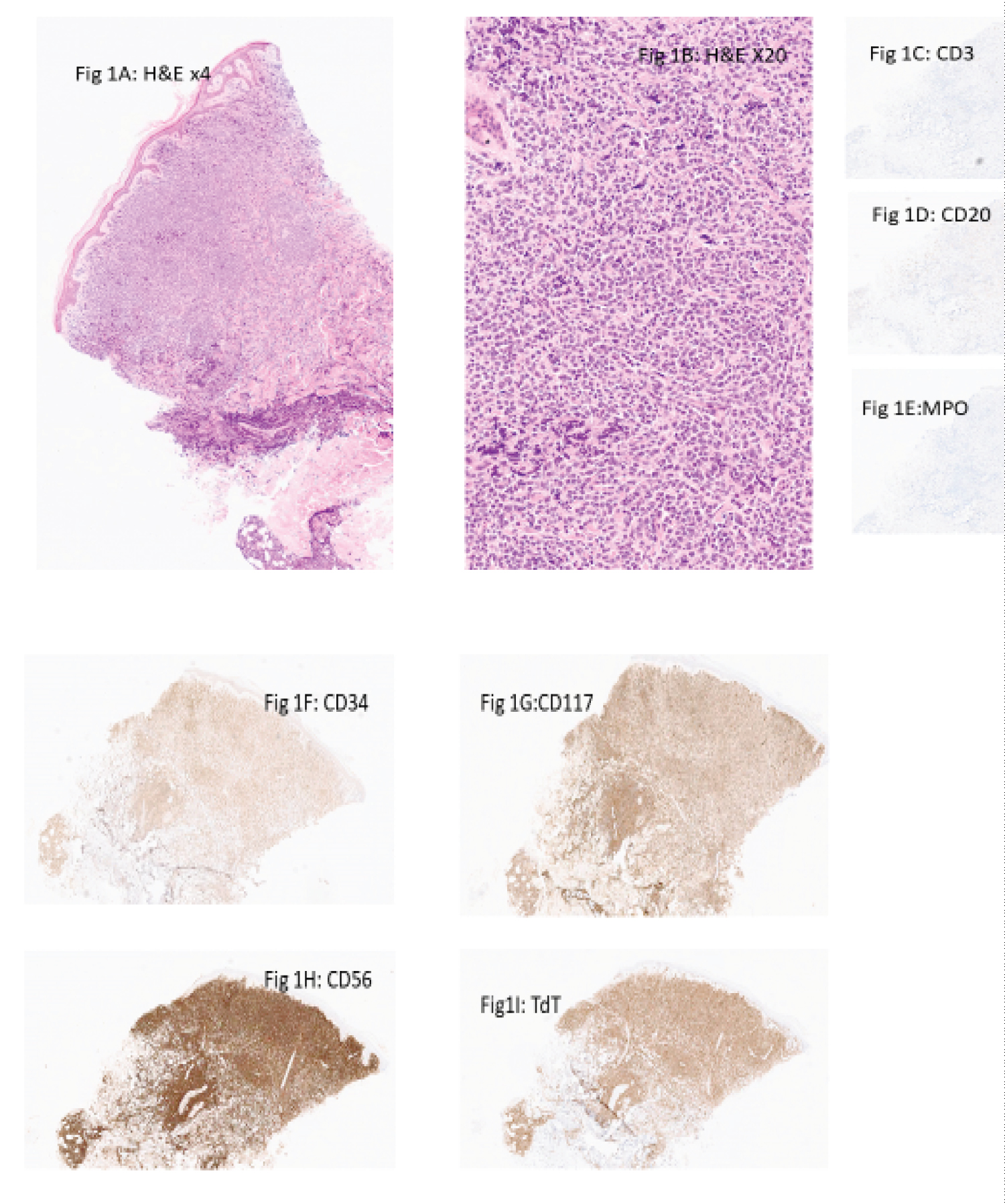 Figure 1: (A&B) show the H&E of the biopsy from the anterior abdominal wall on low and high power, respectively, with negative expressions of CD3, CD20, MPO and positive expressions of CD34, CD117, CD57 and TdT (C-I).
View Figure 1
Figure 1: (A&B) show the H&E of the biopsy from the anterior abdominal wall on low and high power, respectively, with negative expressions of CD3, CD20, MPO and positive expressions of CD34, CD117, CD57 and TdT (C-I).
View Figure 1
Acute leukemia of mixed, or ambiguous lineage, are uncommon, accounting for only 1% of all acute leukemias. Extramedullary presentation is uncommon [1]. The recent WHO classification of Hematolymphoid neoplasms grouped acute leukemia of ambiguous lineage and mixed phenotype acute leukaemia together, and referred them as acute leukemia of mixed or ambiguous lineage (ALAL/MPAL). These either show no evidence of myeloid, B-, or T-lymphoid lineage commitment or show evidence of commitment to more than 1 lineage. This heterogeneous entity is further divided into ALAL/MPAL, with defining genetic abnormalities and ALAL/MPAL that are immunophenotypically defined [2]. Acute undifferentiated leukemia is a subset of the ALAL/MPAL that are immunophenotypically defined. They lack lineage-specific markers, and show expression of no or only one myeloid leukaemia-related immune marker. In other words, they lack B lineage markers (CD19, CD22, and CD79a), T cell lineage markers (cytoplasmic or surface CD3), myeloid markers (MPO) or monocytic markers (non-specific esterase, CD11c, CD14, CD64 or lysozyme). They may express stem cell markers, including CD34, HLA-DR, and terminal deoxynucleotidyl transferase (TdT) [2,3].
Our patient presented with multiple regions of lymphadenopathy and skin nodules showing extensive infiltrates by leukemic cells in which the lineage could not be elucidated. Our differential diagnoses were acute myeloid leukemia with minimal differentiation (AML with minimal differentiation), early T-lymphoblastic leukemia or an aggressive natural killer cell leukemia.
AML with minimal differentiation, like acute undifferentiated leukemia, lacks morphological or cytochemical evidence of myeloid differentiation. MPO is often negative by cytochemistry but may be positive in some blasts by flow cytometry or immunohistochemistry. Blasts of AML with minimal differentiation have at least two myeloid-associated markers, usually CD13, CD117, and CD33. Acute undifferentiated leukemia, alternatively, has zero or only one myeloid-associated marker [3].
CD43 and TdT positivity with other T cell markers being negative may suggest an early T cell differentiation [4].
Our case had a strong CD56 expression. The expression of stem cell markers, with absence of EBER-ISH expression, excluded aggressive natural killer cell leukemia.CD56 is a marker for natural killer (NK) cells, but is also expressed in neoplastic myeloid, lymphoid, plasmacytoid dendritic and myeloma cells, as well as in a minority of T cells. CD56 is also known as neural cell-adhesion molecule (NCAM), which has been associated with shorter remission duration and overall survival, as well as higher cumulative incidences of relapse in acute myeloid leukemia and acute lymphoblastic leukemia [5,6]. The absence of EBER-ISH expression excluded aggressive natural killer cell leukemia.
No firm conclusion has been reached over which induction and consolidation chemotherapy-an AML-based regimen or an ALL-based regimen-should be administered to patients with AUL. Alternatively, most clinicians agree with the necessity of receiving allogeneic hematopoietic stem cell transplantation (Allo-HSCT) to achieve long-term survival because of their poor prognosis [7].
In conclusion, we report a rare case of acute undifferentiated leukemia, localized to the skin and lymph nodes. Exploration of the genomic and epigenetic landscape of acute undifferentiated leukemia will help understand the biologic behavior and ultimately guide therapeutic options.