Seborrheic keratoses (SK) are very common benign epidermal tumors. Their pathogenesis has been detected already in 2006 and includes several aetiological factors. The participation of human papilloma virus (HPV) is being discussed. SK of the penis is extremely rare and may be misdiagnosed. Histopathology will help in the diagnosis.
Seborrheic keratoses, Penis, Human papilloma virus, Histopathological examination
SK: Seborrheic Keratoses; HPV: Human Papilloma Virus
Seborrheic keratoses (SK) are very common benign epidermal tumors. Their pathogenesis has been detected already in 2006 and includes several aetiological factors. The participation of human papilloma virus (HPV) is being discussed [1,2]. SK in the genital area, especially on the penis, is extremely rare and may be misdiagnosed [3]. A careful histopathological examination is essential to establish the correct diagnosis [4].
We report a case of a 50-years-old man, with no significant past medical history and a normal prior sex activity. He presented with a 4-year history of pigmented asymptomatic lesions of the penis. The clinical examination revealed two brown and well demarcated plaques at the base of the penis, measuring 1 × 2 cm and 1 × 1.5 cm, with papillomatous and verrucous surface. The dermoscopic examination showed crypts and brain-like appearance (Figure 1). There was no evidence of lymph node enlargement and the rest of the clinical examination was normal. The patient was deeply frustrated because of the absence of sexual intercourse with his wife since the manifestation of the lesions. The diagnosis of condyloma acuminata, Bowen disease, basal cell carcinoma, melanoma and SK were evoked.
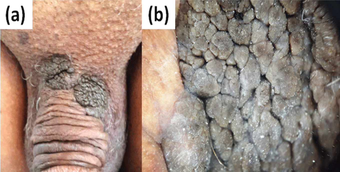 Figure 1: a) Clinical photo: Two brown and well demarcated lesions on the base of the penis, measuring 1 × 2 cm and 1 × 1.5 cm, with papillomatous and verrucous surface; b) Dermoscopic photo: Crypts and brain-like appearance. View Figure 1
The complete hemogram, liver, renal functions and immune status were found to be normal. An examination of human immunodeficiency virus, syphilis, hepatitis B, and hepatitis C proved negative.
The patient was taken to shaving excision of the lesion under local anesthesia. The histopathological examination of the lesions showed hyperkeratosis, papillomatosis and acanthosis with proliferation of basaloid cells containing multiple horn cysts (Figure 2).
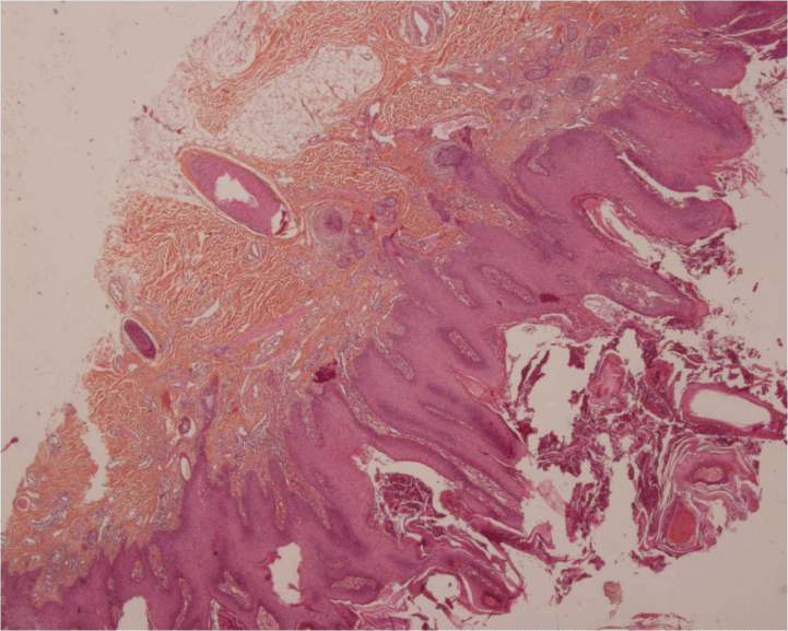 Figure 2: Histological photo: (hematoxylin-eosin, original magnifications × 100). Acanthosis, papillomatosis and horn cysts in the epidermis, with a moderate mononuclear inflammatory infiltrate in the dermis. View Figure 2
Based on the clinical, dermoscopic and histopathological findings, a diagnosis of SK was made. No recurrence was observed during a 15 - month follow-up.
SK are very common epidermal tumors of benign origin. The pathogenesis has been detected already in 2006 but is still not completely understood. Sun exposure, skin phenotype I and II, family predisposition and the activation of the FGFR3 (fibroblast growth factor receptor 3) signaling pathway would be the most likely aetiological factors [1,2].
Recently, a study reported overall mutation rate and also the most frequent mutations through exon sequencing on DNA from SK [5].
It has been suggested that HPV could play a role in the pathogenesis of SK, but he causal relationship is controversial [2]. Tardío, et al. studied the relationship between genital and nongenital SK and HPVs. They confirmed HPVs in 70% of SK in the genital area, whereas in nongenital area, it was only in 5% of cases [6]. Containing HPV 6 low-risk virus, they never lead to malignant transformation.
SK usually occur in patients over 50 years of age. It presents with multiple pigmented papules and plaques with a "stuck on" appearance. Lesions are rarely more than 3 cm in diameter and occur most often on the trunk, face, and extremities, particularly on sun exposed areas. The lesions tend to increase in number and size with advancing age [3]. Morphological variants of SK include the common flat type, skin tag like, stucco keratosis, dermatosis papulosa nigra, inverted follicular keratosis, and melanoacanthoma [7]. The genital region is very rarely affected [3]. The diagnosis of SK can be sometimes challenging, especially in young individuals with lesions in the genital region, and it may be misdiagnosed as condyloma acuminatum, Bowen disease, acrochordons, basal cell carcinoma, and melanoma [3].
The histopathological examination is used to confirm the diagnosis. The typical histopathological findings include acanthosis, papillomatosis, hyperkeratosis, horn cysts, and pseudocysts [8].
The treatment of SK is not mandatory; given the benign origin of the disorder. The most common treatment modalities are cryotherapy, curettage, laser or surgical shave, and other ablative methods [9].
SK in genital area might negatively influence the sexual life of the patient and lead to psychiatric disorders, depression, anxiety, and sexual dysfunction [10].
The penis is rarely affected by SK. This rare condition should be considered in the differential diagnosis for the lesions of the penis and histopathology after shave excision will help in the diagnosis.
None.