Bowen's disease is a squamous cell carcinoma in situ that can degenerate into an invasive squamous cell carcinoma. Early diagnosis and treatment are crucial. There are several therapeutic options available but the most successful is surgical excision with a safety margin. We present a case report of an atypical Bowen's disease in an uncommon location, for which was performed surgical excision with 1cm of safety margin and then the secondary defect was covered by a skin graft. No complications were registered. An acceptable functional and aesthetic outcome was obtained without evidence of relapse.
Bowen's disease, Squamous cell carcinoma, Skin cancer, Safety margin, Surgical excision, Skin graft
Bowen's disease is an in situ squamous cell carcinoma that was firstly described in 1912 by JT Bowen [1]. It is more common in male and caucasians with a peak incidence in seventh decade of life [1].
It usually manifests like an erythematous scaly patch or plaque with irregular borders. However, it can be presented as a verrucous or pigmented lesion [2].
There are several types of treatment described in the literature, such as surgical excision of the lesion with a safety margin, curettage and electrocautery, cryotherapy, laser, photodynamic therapy, topical imiquimod 5% and topical 5-fluorouracil 5%. There is no consensus about the most appropriate treatment [1].
A case of an atypical Bowen's disease in the thenar eminence will be described. This type of cancer is not common in this location and usually doesn't appear like a verrucous lesion. A brief review of this thematic will be done.
The patient is a 41 years old male, skin phototype III of the Fitzpatrick scale, right-hand-dominant with a history of idiopathic chronic renal insufficiency and kidney transplant and Bowen's disease in the left hand submitted to surgery in 2012.
He presented to our department in April 2018 with a verrucous lesion in the right thenar eminence, about 15mm of diameter that had increased in size during the past two years. He was submitted to cryotherapy with a partial response and an incisional biopsy confirmed Bowen's disease (Figure 1).
 igure 1: Preoperative view: presence of Bowen's disease in the thenar eminence.
View Figure 1
igure 1: Preoperative view: presence of Bowen's disease in the thenar eminence.
View Figure 1
His neurovascular exam was intact. He had no palpable lymphadenopathy. Laboratorial analysis revealed no relevant changes.
The treatment was local excision of the lesion with a 1 cm margin of healthy tissue (Figure 2) and the secondary defect was covered by a full thickness skin graft harvested from the internal surface of the right forearm (Figure 3), under general anesthesia.
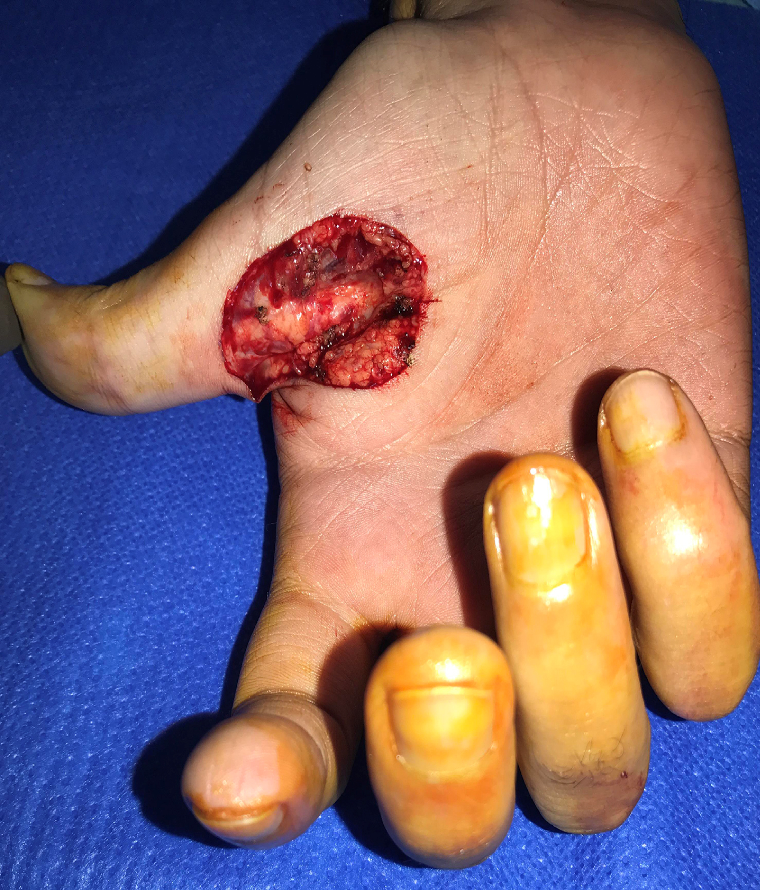 Figure 2: Local excision of the tumor with a safety margin.
View Figure 2
Figure 2: Local excision of the tumor with a safety margin.
View Figure 2
 Figure 3: Intraoperative view: reconstruction of the secondary defect with a full thickness skin graft.
View Figure 3
Figure 3: Intraoperative view: reconstruction of the secondary defect with a full thickness skin graft.
View Figure 3
No lymph node dissection was carried out.
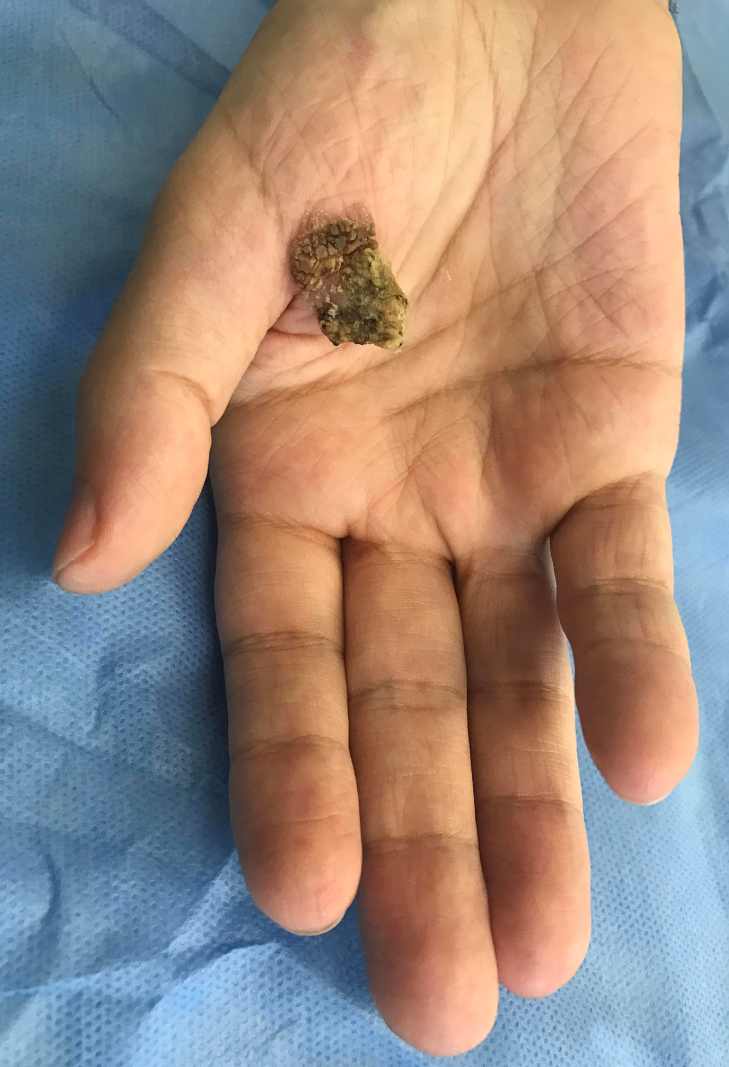
The anatomophatological examination of the lesion (Figure 4) revealed Bowen's disease with free margins.
 Figure 4: Macroscopic view of the tumor.
View Figure 4
Figure 4: Macroscopic view of the tumor.
View Figure 4
There was no postoperative complications.
At 1 month postoperative was achieved an acceptable functional and aesthetic outcome.
Bowen's disease is an uncommon skin disorder. It can appear anywhere on the skin but is specially common on sun exposed areas like head, neck and lower limbs. Although it can arise in genitals, dorsum of the hand and subungueal but is very unusual in palms. Kossard, et al. [3] analysed 1001 cases of Bowen's disease in 1992 and found no cases on the palm. Only a few cases in this location have been reported worldwide.
Clinically, a typical Bowen's disease is a slowly enlarging erythematous patch or plaque, asymptomatic, with 3-5% risk to develop into invasive squamous cell carcinoma in extragenital lesions and about 10% in genital lesions [2]. In most cases it presents as a solitary lesion but in 10-20% of patients multiple lesions may develop [1].
The exact cause is unclear but several etiological factors have been described, such as ultraviolet radiation, radiotherapy, carcinogens (for example arsenic), immunosuppression and viruses like HPV [2].
A diagnosis of Bowen's disease is suspected based upon a detailed patient history and physical examination and is confirmed by a biopsy.
There is a wide range of therapeutic options available for the treatment of Bowen's disease. The selection of the treatment is based on a number of factors such as location of the lesion, diameter, number of lesions, age, comorbidities, immune status and compliance.
Surgical excision is accepted as the gold standard for the treatment of Bowen's disease. It allows to histologically examine the resection margins and ensure free margins.
Depending on size of the secondary defect, it can be closed directly or covered by a skin graft or a local or even free flap.
Non-invasive therapies must be considered in large tumors, multiple lesions or when a poor or delayed wound healing is expected [4].
It is essential an early diagnosis and a prompt treatment due to the risk of malignant transformation. Surgical excision with a wide margin is a useful approach with a low recurrence rate and usually with an acceptable functional and aesthetic outcomes.
There are no conflicts of interest. There are no sources of funding in this paper.