Background: Psoriasis is a fairly common chronic skin condition that presents itself as erythematous, itchy patches with the most commonly affected areas being the elbows, knees, buttocks, scalp, and sites of trauma. Psoriasis of the genitals affects less than half of those diagnosed with psoriasis and can cause major discomfort and frustration. This case report aims to highlight as well as to help discern the presence of vulvar psoriasis among other causes.
Case presentation: This report describes a patient with chronic psoriasis that experienced itching on a skin fold of her genital area. After eliminating other common causes of the symptoms, she was experiencing, such as sexually transmitted diseases, it was determined that the diagnosis was most likely vulvar psoriasis. It was recommended to the patient to wear comfortable, non-irritating underwear, to keep the area well-moisturized with astroglide, and to use a medium-potency steroid ointment. The patient followed-up within a few days stating that she felt much better.
Conclusion: Psoriasis located on the vulva can be difficult to diagnose due to symptoms that can often be confused with other clinical diagnoses. Careful examination and a thorough analysis of the patient's history will lead to the correct diagnosis.
Vulvar presentation, Chronic, Steroid, Genital, Plaque
Involvement of the genitalia occurs in 30-40% of patients with psoriasis, with 2-5% of patients with limited genital psoriasis. A slight increase of prevalence is observed in men vs. women with genital psoriasis [1]. Genital involvement can be associated with considerable psychologic stress, with a negative effect on quality of life, especially on sexual relationships. The resulting psychologic burden makes psoriasis patients more reserved to report genital involvement [2].
Vulvar psoriasis may present as the classic plaque type or inverse psoriasis, with a nonspecific, poorly demarcated erythema. Generally, this presentation also includes mild to severe pruritus of the affected area. Scales of the affected area can be difficult to appreciate in this specific location. A frequent site of involvement is the pubic region, where typical scaling, and/or erythematous macules can be appreciable. In addition, plaques in the pubic region are often denser than in other genital areas and have the potential to coalesce into larger lesions on the labia majora. On the internal lamina of the labia majora, labia minora, and on the clitoris, the lesions are generally more erythematous with few or no scales. The genital area tends to be more prone to friction and perspiration which may cause increased pain or burning sensation due to increased inflammation [3,4].
Genital psoriasis may or may not occur as part of a general psoriasis diagnosis and is not due to any kind of sexually transmitted infection nor is it contagious. Steroid creams or ointments are often used to treat genital psoriasis since typical psoriasis treatments used in other areas are too harsh for the genital skin. The social and economic impact of psoriasis is often underestimated by physicians and health-care providers, with psoriasis affecting the vulvar region providing another layer of difficulty and discomfort as it also becomes a gynecological issue. Patients that experience psoriasis in the genital regions experience significantly worse quality of life than patients with psoriasis in any other areas [5]. This case report aims to provide insight and information on the diagnosis and discernment of vulvar psoriasis.
A 39-year-old reported a feeling of ceaseless itching, specifically in her fourchette. The patient was not sexually active and has been previously diagnosed with psoriasis located on her elbows, hairline, and feet, shown in Figure 1 and Figure 2 below.
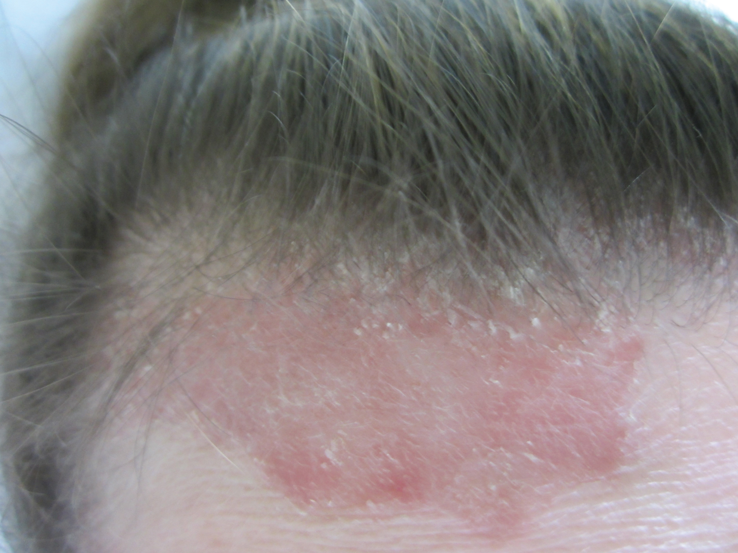 Figure 1: Clinical photograph of erythematous psoriatic lesion located on patient's forehead.
View Figure 1
Figure 1: Clinical photograph of erythematous psoriatic lesion located on patient's forehead.
View Figure 1
 Figure 2: Clinical photograph of multiple crusted scaly psoriatic lesions located on the dorsum of the patient's foot.
View Figure 2
Figure 2: Clinical photograph of multiple crusted scaly psoriatic lesions located on the dorsum of the patient's foot.
View Figure 2
She had frequent recurrences and her treatments involved high potency steroids, both topically and orally, skin moisturizers, coal tar, vitamin D, and ultraviolet rays targeted at her lesions. Her dermatologist referred her back to her primary care physician who then tested her for bacterial vaginosis, candidiasis, and common STDs. All came back negative. During examination a 1.7 cm well-demarcated area in the fourchette between the mucosa and skin was noticed to be erythematous, slightly raised, and non-scaly, presented in Figure 3 below. The patient then identified this as the area that was itching.
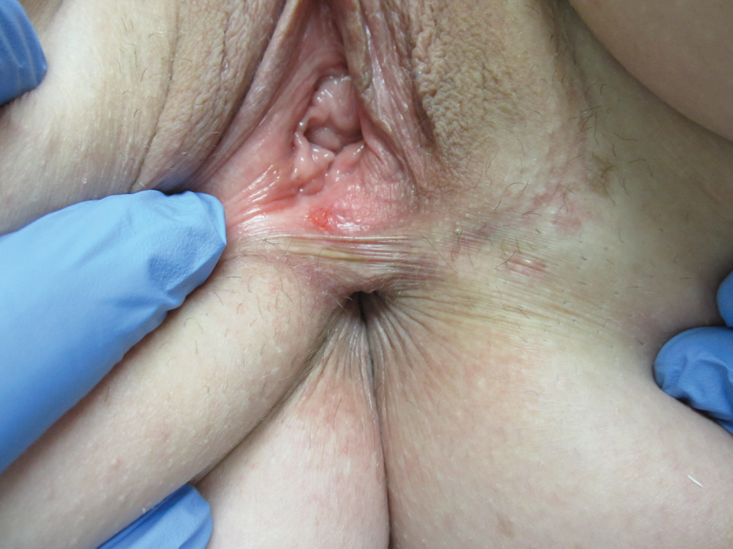 Figure 3: Clinical photograph of a small erythematous, non-scaly psoriatic lesion located in the vaginal fourchette.
View Figure 3
Figure 3: Clinical photograph of a small erythematous, non-scaly psoriatic lesion located in the vaginal fourchette.
View Figure 3
Given her medical history and the clinical presentation of the lesion, the patient was diagnosed with vulvar psoriasis with a good degree of certainty. A biopsy was declined by the patient. The patient was advised to wear smooth cotton unscented underwear, to keep the area well moisturized with Astroglide, and to use a medium potency steroid ointment. She informed our nurse a few days later stating that her condition improved significantly. Similar to her general psoriasis, she was instructed that the rash on her vulvar site will also reoccur from time to time. The patient came into the clinic a few days later reporting that she was feeling much better, and thanked the staff involved.
Diagnosis is based on clinical presentation. Routine biopsy of lesions is not needed for diagnosis, but histopathologic features of vulvar psoriasis are similar to those from non-genital psoriasis. Clinical confirmation is often provided by whole-body skin examination, which reveals features of generalized or inverse psoriasis [6]. Diagnosis may be complicated by superimposed infection, contact dermatitis, or changes of lichen simplex chronicus. Differential diagnosis includes but is not limited to vulvovaginal candidiasis, sexually transmitted infectious, extra-mammary Paget's disease, contact dermatitis, lichen sclerosus, and lichen simplex chronicus. Though there are numerous similarities among the clinical presentations of these conditions, key differences in precipitating factors, etiologies, and physical exam aid in proper diagnosis.
The differential diagnosis of vulvitis can be divided into conditions of infectious and noninfectious etiology. Infectious causes of vulvar pruritis include vulvovaginal candidiasis and sexually transmitted infections. Findings on physical exam, including clumping white discharge in vulvovaginal candidiasis and distinct discharge and lesions in sexually transmitted infections are important distinguishing factors among these conditions [7]. In addition to findings on history and physical exam, analysis of vaginal pH, saline microscopy, and 10% KOH whiff test and microscopy can exclude these infectious diagnoses [7].
The numerous noninfectious and inflammatory causes of vulvar pruritis include extra-mammary Paget's disease, contact dermatitis, lichen sclerosus, lichen simplex chronicus, and vulvar psoriasis. Extramammary Paget's disease is a rare malignancy that can cause vulvar pain and pruritis. The pathogenesis of this disease is not well-defined; however, it is known to affect the sites of apocrine glands. This condition is more common in women and generally affects postmenopausal patients [8]. While the symptoms of extramammary Paget's disease can mimic other inflammatory dermatoses, including psoriasis, the finding of a red plaque with very distinct white scale is morphologically unique to this condition [8].
Contact dermatitis is a noninfectious cause of vulvar pruritis that can occur due to irritation or due to an allergic etiology. While this dermatitis can present with the pain and pruritis seen in vulvar psoriasis, this specific condition is marked by pain due to contact with an irritant, including topical agents or hygiene products. Allergic contact dermatitis also results in pain accompanied by vesicular rash; however, this subtype of contact dermatitis is a delayed hypersensitivity reaction, occurring up to 72 hours after exposure to the allergen [9]. Lichen sclerosus is an inflammatory disorder that can present similarly to contact dermatitis with vulvar pruritis; however, incidence and physical findings in this condition are unique. Lichen sclerosus has a bimodal incidence, with the majority of cases occurring in postmenopausal patients [9]. Additionally, upon physical exam, the skin affected by lichen sclerosus is thin and prone to purpura. This clinical finding is usually identified late in the disease course and is often referred to as a “cigarette paper” appearance [10].
Lichen simplex chronicus, another lichenoid disorder, is an important differential diagnosis that must be carefully distinguished from vulvar psoriasis. This atopic condition is associated with many of the same precipitating factors implicated in other vulvar dermatoses, including exposure to topical irritants and humidity [10]. However, a distinct finding in lichen simplex chronicus on physical exam is vulvar keratinization with erythematous plaques with or without scale [11]. The plaque appearance of this condition can mimic vulvar psoriasis; therefore, thorough consideration of a patient's history and physical exam are necessary to distinguish these conditions. A past history of psoriasis and clinical findings of psoriasis in other locations, as seen in the patient in this case report, support a diagnosis of vulvar psoriasis [11]. If clinical diagnosis is unclear or lesions are unresponsive to treatment, then skin biopsy may be warranted [4,12].
Treatment guidelines for vulvar psoriasis generally follow the same treatment plan as intertriginous psoriasis treatment. First line treatment includes topical steroids. Topical steroid use comes with the risks of atrophy, telangiectasia, striae, and ulceration. Topical vitamin D analogs or topical calcineurin inhibitors are a good option for long term therapy, as they do not share similar risk profiles. Any concomitant infection should be treated accordingly [4,13]. Adjunct treatments include hydration and emollients as keeping psoriatic skin soft and moist minimizes the symptoms of itching and tenderness. Severe vulvar psoriasis may require systemic therapy, such as methotrexate or other biologic agents [14,15].
Vulvar psoriasis can be a difficult diagnosis that should be considered in patients presenting with a chronic erythematous vulvitis without vaginitis. A thorough review of a patient's history and a physical examination should be taken into consideration in order to exclude other common causes of vulvar pruritis. This condition can cause major psychological burden for patients by decreasing one's quality of life and by increasing stress on sexual relationships. Psoriasis and its satellite lesions are a chronic relapsing skin condition that requires long-term management.
We appreciate the Texas Tech University Health Sciences Center, Permian Basin, TX, and Kushal Gandhi, Asley Sanchez, and John Garza at the TTUHSC-PB Research Lab for their assistance and revising with this case report. The authors declare no conflict of interest.
An informed written consent was obtained from the patient.
This research did not receive any specific grant from funding agencies in the public, commercial, or not-for-profit sectors.
GV was in charge of the patient's care and obtained the patient's consent. All authors helped to design the case report. MC and NM contributed equally in writing and editing the manuscript. MS worked extensively on the discussion. All authors have approved a final version of the case report.