A uranyl acetate replacement staining protocol has eliminated the need for highly toxic uranyl acetate in the electron microscopy laboratory. However, the method results in undesirable charging effects when viewed under the transmission electron microscope and can be further improved. A modification of the uranyl acetate replacement staining protocol that simplifies the process is described. The modified method takes less time, and produces better contrast and less charging effects compared to the existing uranyl acetate replacement staining protocol.
Uranyl acetate, Modified uranyl acetate replacement, Staining protocol, Transmission electron microscopy, Samarium triacetate, Gadolinium triacetate
In electron microscopy, images are really no more than magnified projections of the various densities proportionate to the components of the section. In order to achieve a differential increase of the densities in biological structures, differential contrast is needed to produce a sharp image definition.
In transmission electron microscopy (TEM), contrast is mainly produced by electron scattering at the specimen. Structures that strongly scatter electrons appear as dark areas while structures that scatter fewer electrons appear bright [1]. To increase their contrast, electron dense stains can be added to the sample. Staining is usually done with heavy metal salts commonly derived from uranium, tungsten and lead. Heavy metal ions are used since they will readily interact with the electron beam and produce phase contrast [2].
The most frequently used method for staining in TEM is a two-step procedure of staining with Uranyl Acetate (UA), followed by lead citrate [3]. Although UA is an excellent and well-characterized stain, replacements are sought for several reasons. When it needs to be handled as a powder, it is very toxic and carcinogenic if inhaled. Furthermore, depleted UA is considered a radioactive material, and hence subjected to stringent regulations by the authorities. Therefore, UA requires safe storage and careful handling, which in turn increases cost for shipping and waste disposal.
In 2011, two lanthanide salts, samarium triacetate and gadolinium triacetate, were found as staining reagent substitutes for UA [4]. Lanthanide salts work the same way as heavy metal salts. They will stain certain biological components such as proteins and phospholipids, and interact with electron beams to produce contrast. These salts were called Uranyl Acetate Replacement (UAR). UAR is considered safer as there is no radioactive material used and can be purchased from Electron Microscopy Sciences, USA. In our preliminary study, UAR staining protocol with mouse kidney ultra-thin sections produced charging effects and reduced contrast and sharpness. Charging effects cause electrons to accumulate during staining and affect the quality of the stained specimen.
In this study, we would like to examine the effect of adding a simple modification step to UAR. We called this Modified Uranyl Acetate Replacement (MUAR), which is the addition of a post-staining step using lead citrate. It was hypothesized that this step will reduce the charging effect and increase the contrast and sharpness of the electron micrograph from TEM.
We manually stained mice kidney tissue samples using the staining protocols of UA, UAR and MUAR.
The mice were sourced from Laboratory Animal Resource Unit, Institute for Medical Research. Freshly dissected mouse kidney tissues were fixed in 2.5% phosphate buffered glutaraldehyde and post-fixed with 2% osmium tetroxide. Mice kidney tissues were dehydrated with 50%, 70%, 90% and 100% (3X) acetone and embedded in Agar 100 epoxy resin and sectioned to a 90 nm thickness. A sample grid is floated, sectioned side down on the stains following the protocols of UA, UAR and MUAR (Table 1). After blotting off the stains, the grid was rinsed thoroughly with double-distilled water to remove any residual unbound stain. The protocols of UA and MUAR required second staining using lead citrate and washed as before. The stained ultra-thin sections of mouse kidney were viewed under TEM (FEI Tecnai G2) at 100 kW.
Table 1: Details of Manual Staining Protocols. View Table 1
A mixture of 1.33 g lead nitrate and 1.76 g sodium citrate were shaken vigorously in 30 mL distilled water. The dissolved solution appeared milky white in colour. One millilitre of 1 N sodium hydroxide was added and mixed well until the solution turned clear. Finally, the solution was topped up to 50 mL using distilled water and the pH adjusted to 12.
One gram of Uranyl Acetate was dissolved in 50 mL distilled water.
Uranyl Acetate Replacement was purchased from Electron Microscopy Sciences, USA. The preparations of the UAR and MUAR stains are as shown in Table 1.
Figure 1 shows the TEM images obtained using the different staining protocols as described in Table 1. The three images show variation in the degree of sharpness, contrast and brightness in the stained kidney sections. The MUAR protocol produced more detailed lines of structures that can be clearly seen compared to other protocols. Differential contrasts between the different biological structures were well defined in MUAR protocol than in other protocols. There were significantly more charging effects on the kidney sections in UAR compared to other protocols. Sharpness, contrast and brightness between the MUAR and UA protocol were of high quality in MUAR compared to UA.
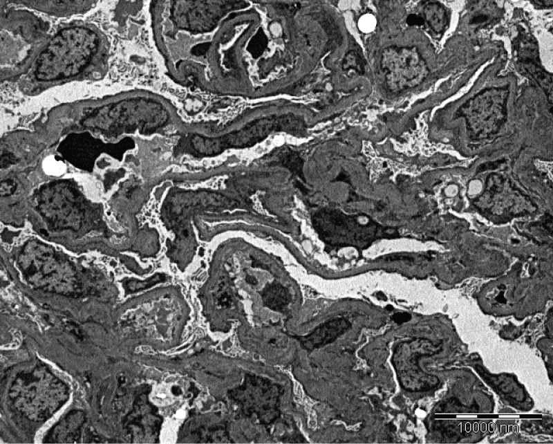 Figure 1A: Mice kidney section stained using MUAR protocol. Sharpness, contrast and brightness were well defined using MUAR compared to UAR and UA. Magnification at 4500X and scale bar is 10,000 nm. View Figure 1A
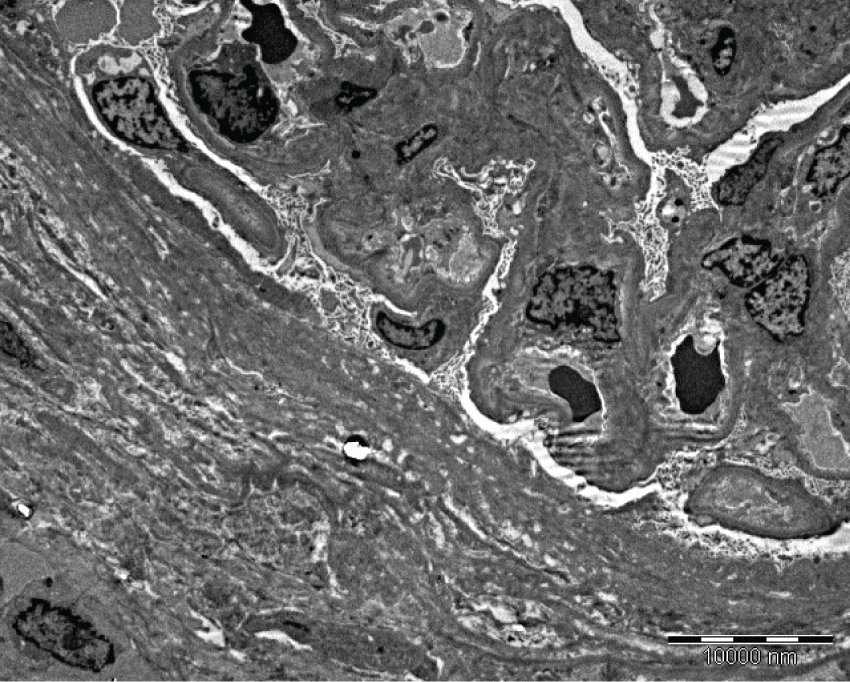 Figure 1B: Mice kidney section stained using UA protocol. Sharpness, contrast and brightness using UA protocol were less defined than MUAR but better than UAR. Magnification at 4500X and scale bar is 10,000 nm. View Figure 1B
The staining protocol in MUAR is superior compared with UAR and UA. MUAR produces quality kidney images and provides defined ultra structures of the kidney's organelles. Moreover, MUAR needs shorter time to complete compared to other staining protocols (Table 1). The total protocol time for staining kidney sections is less than 15 minutes including the washing steps. MUAR staining protocol provides an alternative staining to the UA staining (which is the gold standard in staining electron microscopy ultra-thin sections) and it is safer to use as did not contain any radioactive substances. The MUAR protocol has been used in our laboratory for about a year. Apart from kidney, other cell cultures and organ tissues such as heart, muscle, and liver have been examined successfully using MUAR staining protocol in our laboratory.
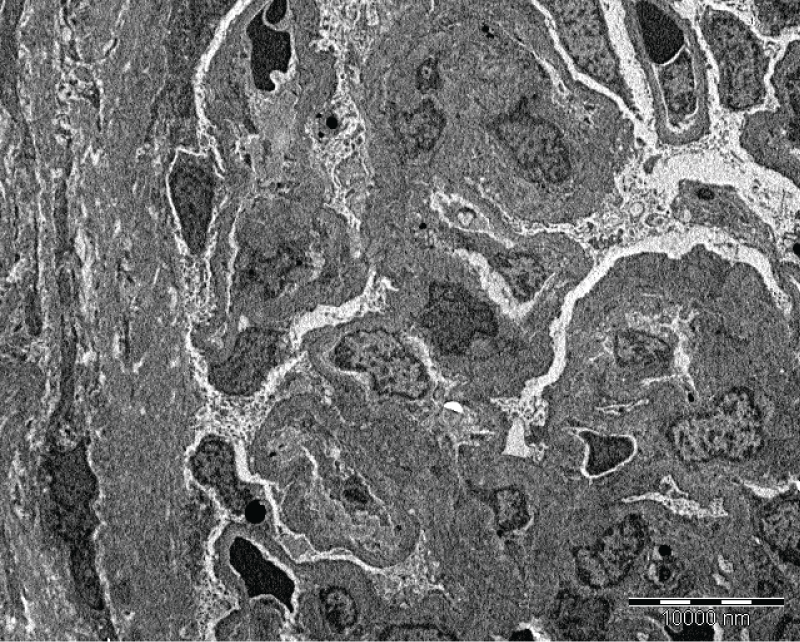 Figure 1C: Mice kidney section stained using UAR protocol. Charging effect were very obvious compared to MUAR and UA. Magnification at 4500X and scale bar is 10,000 nm. View Figure 1C
Besides lanthanide salts, other staining reagents have been developed for TEM. One of them is oolong tea extract (OTE). The polyphenolic components in OTE are thought to react with peptide bonds. The contrast produced by reaction with OTE can be further enhanced with lead citrate stain [5]. Another alternative stain to UA is a compound called Platinum Blue (Pt Blue), which is a product of the reaction of cis-dichloro-diamine-platinum (II) with thymidine [6]. Although Pt Blue can produce better contrast than OTE, Pt Blue is hazardous while OTE is harmless for health and environment. However, we have not tested both of these staining reagents in our laboratory that enable us to make any direct comparison.
In the past decade, alternative staining reagents such as UAR, OTE and Pt Blue have been developed to replace UA in electron microscopy. The present study showed that a simple modification to UAR protocol, by adding a second lead citrate staining step, can increase the contrast and image quality of TEM. This MUAR staining protocol is safe, faster and can easily be adopted by other laboratories.