Introduction: To demonstrate the contribution of the multiple uniport passages technique without co-axial during ultrasound-guided biopsy sampling of breast masses in interventional radiology settings in Kinshasa, Kinshasa University clinics in particular.
Materials and Methods: This was a retrospective, descriptive, multicenter study conducted over a 6-year period from June 2018 to June 2024 and compiled at the university clinics of Kinshasa. Any patient who underwent percutaneous echo-guided biopsy sampling using the multiple-pass technique without co-axial was included. Biopsy specimens were taken using 14 to 17 G tru-cut needles. Data analysis was performed using SPSS version 2.2 software, which enabled us to collect 107 patients.
Results: The mean age of our patients was 43.5 ± 17.5 years. The age of the majority of our patients ranged from 37 to 69 years. The majority of patients were referred for a left breast mass (47%), and 100% of our patients underwent ultrasound guidance during breast biopsy. Automatic guns were used most frequently (88.78%). The technique of multiple uniportal passages without co-axis was conclusive in almost all our patients (100%). Malignant lesions were the most common (37%). Metaphasic carcinoma was the most common histological type, accounting for 18.5% of cases. No major complications were recorded during the various ultrasound-guided breast biopsy punctures.
Conclusion: Percutaneous ultrasound-guided breast biopsy is an interventional radiology procedure; the multiple uniport passages technique without co-axial is an innovative, effective alternative for ultrasound-guided biopsy sampling of breast masses in hospitals in Kinshasa. It has proved effective in almost all our patients, yielding excellent and conclusive cores.
Biopsy puncture, Ultrasound-guided, Co-axial, Multiple passage
A breast biopsy is usually performed after the discovery of a suspicious lesion by mammography or ultrasound, in order to obtain tissue for pathological diagnosis [1]. Several methods of breast biopsy are now available [2]. These include fine needle aspiration (FNA), vacuum-assisted biopsy, trocar biopsy and surgical excision biopsy [3-5]. In addition to ultrasound, breast biopsies can be performed using MRI or stereotactic biopsy imaging guidance [2,4-6]. Vacuum-assisted biopsies are generally performed using stereotactic techniques when the suspicious lesion can only be seen on mammography [5,6]. Needle biopsies have largely replaced open surgical biopsies [7,8]. There are therefore numerous indications for breast biopsies, based on the ACR classification. It should be noted that abnormalities classified as ACR 4 account for the vast majority of these indications. Breast biopsies performed for ACR 5 lesions are part of an approach designed to optimize therapeutic management [7,8]. In practice, ACR 3 poses problems of interpretation. BI-RADS 3 classification is accompanied by a high degree of inter-observer variability. Trocar biopsy or core needle biopsy (CNB) is another percutaneous (“through-the-skin”) method of breast biopsy that became more popular than FNA in the 1990s due to the larger tissue sample provided by CNB [9]. This method is usually performed under ultrasound guidance and involves the use of two needles, an internal “puncture” needle that is inserted into the mass, and a larger-gauge needle with an open “gap” or “hollow” on one side that allows tissue to penetrate [10-14]. A spring-loaded sheath is then triggered by the technician, covering the hollow of the needle to allow the tissue to be separated and removed from the sample for analysis [11-14]. As a rule, four tissue samples are taken to minimize sampling errors. To avoid having to pierce the breast repeatedly, a coaxial needle is left in place above the mass as a guide [12-14]. CNB has a higher sensitivity for cancer than FNA, has fewer false negatives and has proved more effective in finding rare breast diseases such as lobular carcinoma. Percutaneous (“through-the-skin”) biopsy methods have become more preferred than surgical biopsies due to the high rate of benign findings (80%) and the reduction of adverse effects such as scarring [10-14]. Obtaining a histological diagnosis by microbiopsy sampling is essential to define the optimal therapeutic strategy for a breast tumor. Microbiopsy is the technique of choice, as it provides precise tumour type and the main histological prognostic and predictive factors, while being simple, rapid and inexpensive [15-17] and thus allows the indication of pre-operative breast lymphoscintigraphy for infiltrating cancers, with selective sentinel lymphadenectomy at the time of surgery [16-17]. The aim of the present study is to demonstrate the interest and practical modalities of an original method of ultrasound-guided breast microbiopsy using the technique of multiple uniportal passages without co-axial useful in the management of solid or solid-cystic breast tumours in Hospital settings in Kinshasa.
This was a descriptive, multicenter, observational study of the various echoguided percutaneous puncture-biopsy procedures performed using the multiple-pass technique without co-axial during a 5-year period, from January 2018 to June 2023.
We conducted this study in seven hospital institutions in the city and province of Kinshasa, including one tertiary-level institution, the Cliniques Universitaires de Kinshasa. Other institutions included: Centre Médical Diamant de Kinshasa, Clinique Présidentielle de l'Unité Africaine, Centre Hospitalier de Kingasani, Centre d'Imagerie Médicale Pilote Kokolo, Centre Pistis Médical Center de Limete and Vision Médicale pour Tous. All these data were coded at the Cliniques Universitaires de Kinshasa, located on Route de Kimwenza, in the commune of Lemba, district of Mont-Amba, in the city-province of Kinshasa in the Democratic Republic of Congo.
One hundred and seven patients out of a total of six hundred and fifty-six who had undergone various interventional radiology procedures in Kinshasa hospitals were included in the present study. The mean age was 43.5 ± 17.5 years, with extremes ranging from 15 to 81 years. The majority of patients in the series were between 37 and 69 years of age. Of the 107 patients, 5 were male and 102 female.
Any male or female patient referred for ultrasound-guided breast biopsy with a medical imaging result (breast ultrasound, mammography and/or breast MRI); Any patient with a normal haemostatic work-up (normal bleeding time and coagulation time); Any patient with a haemoglobin and/or haematocrit level within acceptable limits (haemoglobin ≥ 10 g/dl and haematocrit ≥ 30%), any patient who has freely consented in writing to the ultrasound-guided breast microbiopsy procedure.
The following were not included in the present study: Any patient referred for ultrasound-guided breast biopsy who did not have a previous breast imaging result; any patient with a severely disturbed haemostatic balance (TS: TC : ); any patient with a low haemoglobin and/or haematocrit level (Hb ≤ 7 g/dl and Hct ≤ 321%) refractory after correction ; any patient who had not freely given written consent for the indicated interventional radiology procedure. On the other hand, patients with low hemoglobin levels (below 7.5 g/l) were presented to the staff for validation of the procedure.
Parameters of interest:
Socio-demographic parameters : Included patient age, sex and place of origin.
Clinical parameters : Included clinical reason for referral for breast biopsy, minor or major incident after biopsy.
Biological parameters : Included hemoglobin level, hematocrit level, red blood cell count, bleeding time, coagulation time, and pathological findings.
Radiological parameters : Included the means of imaging used to perform the biopsy, the indication for breast biopsy, the equipment used for the biopsy (automatic gun, semi-automatic gun, suction gun, not forgetting the drainage trocar); needle insertion techniques (co-axial or indirect technique and axial or direct technique), type of anesthesia used (local anesthesia or narcosis), anesthetic used (lidocaine vs xylocaine without preservative), number of cores obtained, the number of passages performed, whether the biopsy path was embolized or not, whether haemostatic products were used or not, whether the clot was used or not, the type of premedication, the anatomopathological result and finally the sampling technique used was Multiple passages uniport without coaxial (TMPUSC).
Precautions : Oral and written consent was obtained. A haemostasis test was carried out 12 to 48 hours before the procedure. A platelet count greater than or equal to 150,000/mm 3 and a prothrombin time (PT) greater than or equal to 70% were required for the procedure. We used automatic and semi-automatic biopsy needles ranging from 18 to 14 Gauges (G) and 10 to 15 cm in length, depending on availability.
Equipment : After radiological consultation, the examinations were reviewed and the lesion re-evaluated by B-mode ultrasound and duplex Doppler using convex and linear multi-frequency probes. Several brands of ultrasound equipment were used, including Phillips U-22, Sonoscape Light, Sonoscape S-50 and Mindray.
Technique : A manual ultrasound scan was performed prior to any procedure. Rigorous asepsis of the skin planes was obtained using povidone-iodine. After sterile draping, we administered a local subcutaneous anaesthetic at the entry point and along the chosen route to the peri-lesion, with 5 to 10 ml of preservative-free 2% lidocaine. The biopsy trocar was inserted under ultrasound guidance to within 10 mm of the target, with good visualization of the bevel. No coaxial was used, and the technique chosen by our team for sampling was that of multiple passage uniport without coaxial (TMPUSC). Four to eight samples were taken, depending on the calibre of the biopsy trocar used and the patient's compliance. The cores obtained were fixed in formalin 4 or 15% and sent to the pathology laboratory for morphological and immunohistochemical analysis. All breast microbiopsies were performed in the day hospital. Patients were monitored clinically and ultrasonographically, with a breast ultrasound scan performed at the end of the procedure. The patient was placed in the right lateral decubitus position for 40 minutes to an hour before being discharged home. On discharge, painkillers were systematically prescribed, but antibiotic prophylaxis was recommended on a case-by-case basis in consultation with the referring physician.
Over a period of 6 years, we conducted a descriptive, observational, multicenter study of 107 patients who had undergone percutaneous ultrasound-guided puncture-biopsy using the multiple uniport passages without co-axial (MPUSC) technique, in hospitals in Kinshasa. This series enabled us to highlight the following results: Socio-demographic characteristics: mean age 43.5 ± 17.5 years, with extremes ranging from 15 to 81 years (Table 1). The majority of patients in the series were between 37 and 69 years of age. With regard to gender, 95.3% of our patients were female, with a calculated M/F sex ratio of 0.049 (Table 1). Clinical information. The majority of our patients were referred for left breast mass (47%), followed by right breast mass (36%) (Table 2). Histological data: Macroscopic analysis of the various cores revealed a pearly white appearance in all samples taken (Table 3). Malignant lesions accounted for 37% of all lesions in our series, with a predominance of metaphasic carcinomas (18.5%). followed by adenocarcinoma (14.8%) and infiltrating mucinous carcinoma (3.7% of cases) (Table 3, Figure 1). Benign lesions accounted for 29.7% of cases, with fibrocystic mastosis the most common, accounting for 18.5% of all lesions. Fibroadenomas accounted for 7.4% (Table 3, Figure 1). Infectious lesions were found in 5.6% of cases; 3.7% for chronic active mastitis and 1.9% for mammary and lymph node tuberculosis (Table 3, Figure 1). Analysis of correlations between anatomopathological findings and patients' sex revealed a predominance of metaphasic carcinoma in female patients for malignant lesions, and fibrocystic mastosis for benign lesions, respectively at a level of (Table 3). Radiological data: With regard to the guidance used in the present series, it should be noted that 100% of our patients had ultra sonographic guidance during biopsy (Table 4). Haematological and haemostatic tests: these were within normal ranges in all our patients, i.e. 100% of the total (Table 5). The following tests were performed: Bleeding time, coagulation time, hemoglobin level and hematocrit level (Table 4). Concerning pre-procedure preparations: Psychological preparation was performed in all our patients (100%), atropine premedication was administered in all our patients (100%), hemostatic assessment was performed in all our patients (100%) (Table 5). With regard to sampling equipment, automatic guns were the most widely used in the present series, accounting for 88.78% (Table 5). Automatic 14-Gauges guns were the most widely used, while semi-automatic guns were used by 11.21% (Table 5). With regard to the core sampling technique: The uniport multiple-pass technique without co-axial was used in almost all our patients (100%), with successful sampling in almost all our patients (Table 5). No embolization of the biopsy path was performed (Table 5). As for the sampling technique used, it should be noted that the multiple passage uniport without co-axial technique (MPUSC) was used in 100% of our patients (Table 5). Local anesthesia was used in 106 patients (99.06%); only one patient in the present series had benefited from general anesthesia (0.93%) (Table 5). Lidocaine and/or xylocaine without preservatives was used in almost all our patients (100%). Bicarbonate was not used in the present series (Table 5). With regard to complications, no minor, major or fatal complications were encountered in the present series (Table 6).
 Figure 1: Number of cores after sampling.
View Figure 1
Figure 1: Number of cores after sampling.
View Figure 1
Table 1: Distribution of patients by age and sex. View Table 1
Table 2: Distribution of patients based on clinical information. View Table 2
Table 3: Distribution of patients based on anatomopathological results. View Table 3
Table 4: Distribution of patients based on guidance methods used. View Table 4
Table 5: Hemostasis assessment / Preparation before procedure, types of needles, technique used and type of anesthesia. View Table 5
Table 6: Complications. View Table 6
Determining the histological nature of a tumour is important for its management. Despite technological advances in imaging, biopsy sampling is still necessary for anatomopathological and immunohistochemical analyses [3,18]. Biopsy puncture was preferred to cytopuncture in 100% of our patients. This choice was based on the fact that biopsy yields more analyzable tumour material [4,18,19]. The sampling technique chosen was that of multiple uniport passages without co-axial, used in almost all our patients (100%). Our samples were conclusive in all patients (100% of cases). This very satisfactory result can be explained by the fact that all our biopsies were performed under ultrasonographic guidance in order to select not only the target lesions, but also the suspect areas to be sampled [19-21]. In the present series, all our biopsies were performed using the MPUSC technique, which consisted first of all in ultrasound identification of the lesion prior to any procedure. This was followed by rigorous asepsis of the skin planes using povidone-iodine, sterile draping, local subcutaneous anesthesia at the entry point and then all along the chosen route to the peri-lesion. After infiltration of 5 to 10 ml of preservative-free 2% lidocaine, the biopsy gun was introduced under ultrasound guidance, without the co-axial in place; up to 10 mm from the target, with good visualization of the bevel. Four to eight samples were taken, depending on the calibre of the biopsy trocar used and the patient's compliance. The cores were fixed in 4% or 15% formalin and sent to the pathology laboratory for morphological and immunohistochemical analysis. All breast microbiopsies were performed on an outpatient basis [2,18]. Local anaesthesia was used in 106 patients (99.06%); only one patient in the present series had benefited from general anaesthesia (0.93%). The biopsy was performed under general anaesthetic, as the patient was being followed for bilateral ulcerated breast adenocarcinoma at the inoperable stage, in whom we performed a double breast biopsy on the ulcerated breasts. Our study showed that malignant lesions accounted for 37% of all lesions in our series, with a predominance of metaphasic carcinomas (18.5% of cases), followed by adenocarcinomas (14.8% of cases) and infiltrating mucinous carcinoma (3.7% of cases) [8]. These results are in antiphase with those found by Bouaziz [7,8], who reported an adenocarcinoma rate of 90.4%, and Aka [7,8], who found an adenocarcinoma rate of 72.3%. However, we found a similarity with mucinous carcinoma, which remains a rare entity present in 3.7% of cases, in line with the literature, which reports a rate of between 1 and 5% [7]. Benign lesions were found in 29.7% of cases, with fibrocystic mastosis leading the way at 8.5%, followed by fibroadenomas (7.4%). These results contradict those reported in the literature, where 50% of breast biopsies are performed [2,3]. This could be explained by our relatively small cohort compared with other studies. No minor or major complications were noted during the performance of our procedures, although long-term follow-up was not carried out to look for any swarming of the biopsy pathway. This proves the dexterity of the procedure and the technique used by our team. The mean age of our patients was 43.5 ± 17.5 years, with extremes ranging from 15 to 81 years. The majority of our patients were between 37 and 69 years of age. Ninety-five percent of our patients were female, with a calculated M/F sex ratio of 0.049. Our results are similar to those reported by the Ghanaian and Nigerian studies on the subject, which reported a mean age of 49.19 and 48 respectively [8,9]. Our results are close to those in the literature; among others in the USA, 50% of breast cancer cases are diagnosed in women aged over 65 [10]. In Europe, for women aged 50 to 54, the incidence of breast cancer is 210 per 100,000, rising to over 300 per 100,000 women at the age of 70 and to over 430 per 100,000 women for those aged over 80 [7,8]. The rarity of breast tumours in men can be explained by the atrophic nature of the gland, the thinness of the milk ducts, the absence of acini and the abundance of fibrous tissue. Left-breast involvement was predominant in 47% of our patients, versus 36% with right-breast involvement. In 56.32% of cases, the tumour involved the left breast. The predominance of cancer in one breast compared with the other can be explained by breastfeeding habits [12,22]. Automatic guns were most commonly used in 88.78% of our patients versus semi-automatic used in 11.21%. The 14-Gauge size was the most widely used in the present series. The use of automatic and 14 G needles would be dictated by several reasons among others: The availability of materials, the mobility of most breast lesions; not forgetting the good size of cores with 14G, thus enabling anatomopathologists to make better analyses.
Percutaneous puncture biopsy techniques are currently at the forefront of the diagnostic approach in senology. They are an excellent alternative to surgical biopsy. Indications for percutaneous sampling are based on the ACR's BIRADS classification. Abnormalities classified as ACR BIRADS 4 (Figure 2) account for the vast majority of indications. Breast biopsies performed for lesions classified as ACR BIRADS 5 (Figure 3, Figure 4) are increasingly likely to be part of an approach designed to optimize therapeutic management. ACR 3 BIRADS 3 (Figure 5) is the category that poses the most interpretation problems in practice. Nevertheless, in certain cases, percutaneous sampling may be advisable, particularly in cases of personal or family risk factors. In the present study, the mean age of our patients was 43.5 ± 17.5 years. The age of the majority of patients in the present series ranged from 37 to 69 years. The majority of our patients were referred for a left breast mass (47%), and 100% of our patients had benefited from ultra sonographic guidance during breast biopsy. Automatic guns were used in 88.78% of cases, and the technique of multiple uniportal passages without co-axis was successful in almost all our patients (100%) (Figure 6). Malignant lesions accounted for 37% of all lesions. Metaphasic carcinomas were diagnostic in 18.5% of cases. No major complications were recorded during any of the ultrasound-guided breast biopsy procedures.
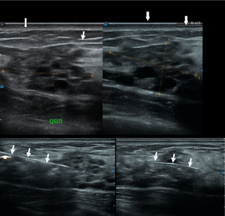 Figure 2: A 34-year-old patient presenting with a pre-pectoral solid lesion measuring 8.58x7.83x5.25 mm in diameter, i.e. an overall volume of 0.18 ml, classified as BIRADS 4 by the ACR, associated with a lesional patch on the left QSI classified as BIRADS 3 by the ACR. The various arrows indicate the masses, while the arrowheads indicate the biopsy needle paths.
View Figure 2
Figure 2: A 34-year-old patient presenting with a pre-pectoral solid lesion measuring 8.58x7.83x5.25 mm in diameter, i.e. an overall volume of 0.18 ml, classified as BIRADS 4 by the ACR, associated with a lesional patch on the left QSI classified as BIRADS 3 by the ACR. The various arrows indicate the masses, while the arrowheads indicate the biopsy needle paths.
View Figure 2
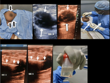 Figure 3: Patient aged 25 years, presenting with a hypoechoic solid mass in the QSE of the left breast classified as BIRADS 4 by the ACR. Black and white arrows: indicate the left breast before biopsy, white arrows: indicate the hypoechoic solid mass of the left QSE, arrowheads: indicate the hyperechogenic image representing the biopsy gun.
View Figure 3
Figure 3: Patient aged 25 years, presenting with a hypoechoic solid mass in the QSE of the left breast classified as BIRADS 4 by the ACR. Black and white arrows: indicate the left breast before biopsy, white arrows: indicate the hypoechoic solid mass of the left QSE, arrowheads: indicate the hyperechogenic image representing the biopsy gun.
View Figure 3
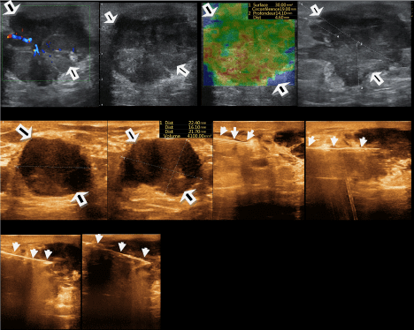 Figure 4: A 53-year-old patient with a solid mass in the QSE of the left breast classified as BIRADS 5 by the ACR, who underwent an ultrasound-guided microbiopsy. Images 1-2-4-5 and 6: are B-mode ultrasound images, showing the solid, hypoechoic, irregularly contoured mass, classified as ACR BIRADS 5. Image 3 is an ultrasound image showing areas of elastographic rigidity. Images 7-8-9-10 are B-mode images, showing the path of the biopsy hyperechoic linear lines.
View Figure 4
Figure 4: A 53-year-old patient with a solid mass in the QSE of the left breast classified as BIRADS 5 by the ACR, who underwent an ultrasound-guided microbiopsy. Images 1-2-4-5 and 6: are B-mode ultrasound images, showing the solid, hypoechoic, irregularly contoured mass, classified as ACR BIRADS 5. Image 3 is an ultrasound image showing areas of elastographic rigidity. Images 7-8-9-10 are B-mode images, showing the path of the biopsy hyperechoic linear lines.
View Figure 4
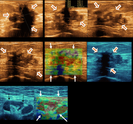 Figure 5: A 62-year-old patient with a solid QS mass classified as ACR BIRADS 5, who underwent an ultrasound-guided microbiopsy. Images 1-2-3-4 and 6: are B-mode ultrasound images, showing a solid, hypoechoic, irregularly contoured, prepectoral mass, classified as ACR BIRADS 5. Images 5 and 8 are elastography images: showing areas of rigidity within the mass. Image 7 shows an axillary adenopathy with a secondary appearance.
View Figure 5
Figure 5: A 62-year-old patient with a solid QS mass classified as ACR BIRADS 5, who underwent an ultrasound-guided microbiopsy. Images 1-2-3-4 and 6: are B-mode ultrasound images, showing a solid, hypoechoic, irregularly contoured, prepectoral mass, classified as ACR BIRADS 5. Images 5 and 8 are elastography images: showing areas of rigidity within the mass. Image 7 shows an axillary adenopathy with a secondary appearance.
View Figure 5
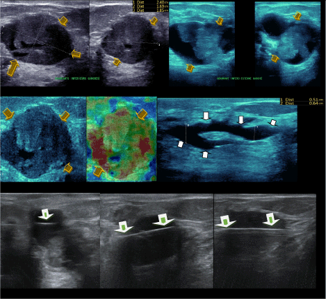 Figure 6: 40-year-old patient with two solid endo-canal masses in the QSE of the left breast and ectasia of the galactophore ducts with évidence of luminal infiltrating Carcinoma B: possibility of HR without HER-2/Neu oncogene over expression. Images 1 to 5: with yellow arrows: represent the two solid endo-canal masses of the left QSE ; image 6: shows areas of stiffness within the mass on elastography the 7 -th image with white arrows: indicate ductal dilatation, images 8-9-10: with white-green arrows: indicate biopsy gun paths.
View Figure 6
Figure 6: 40-year-old patient with two solid endo-canal masses in the QSE of the left breast and ectasia of the galactophore ducts with évidence of luminal infiltrating Carcinoma B: possibility of HR without HER-2/Neu oncogene over expression. Images 1 to 5: with yellow arrows: represent the two solid endo-canal masses of the left QSE ; image 6: shows areas of stiffness within the mass on elastography the 7 -th image with white arrows: indicate ductal dilatation, images 8-9-10: with white-green arrows: indicate biopsy gun paths.
View Figure 6
As a first limitation, we noted: the small sample size, the lack of certain clinical, biological and mammographic information of our patients constitute the limitations of the present study.
Breast cancer is the most common cancer in women; it is particularly increasingly common in developing countries, where the majority of cases are diagnosed at advanced stages; early detection remains the principal means of combating the disease. Breast microbiopsy is an essential step in the diagnosis of breast cancer. Biopsy is essential because the diagnosis of cancer is a histological one. It consists in taking a sample of tissue fragments from the lesion suspected of being cancerous. It is performed by a radiologist under ultrasound guidance.
The present study is descriptive and retrospective, based on our local experience over the last six years. It is a first to our knowledge, highlighting an innovative, simple and effective technique known as the “Technique de multiple passages uniport sans co-axial,” TMPUSC, for echo guided biopsy sampling of breast masses in hospital settings in Kinshasa. It avoids the use of haemostats, abundant bleeding at the puncture site and embolization of the biopsy path
The authors declare that this study contains no personal data likely to identify the patient or subject.
This study has not received specific funding from any public or private institution.
All authors have no possible conflict of interest.
1. Frederick Tshibasu Tshienda: editing and proofreading;
2. Tasnime Hamdeni: editing and proofreading;
3. Najat cherif Idrissi El Ganouni3: editing and proofreading;
4. Michel Lelo Tshikwela: editing and proofreading;
5. Jean Mukaya Tshibola: editing and proofreading;
6. Jean Marie Kayembe Ntumba: editing and proofreading.
7. Jean-Marie Mbuyi Muamba: editing and proofreading.