Objectives: The purpose of this narrative review is to describe effective efforts in radiation protection to limit scattered radiation exposure in terms of room analysis and to find out the difference in the effect of retaining wall thickness in limiting the scatter radiation dose.
Reviews: Scattered radiation is part of radiation that deviates from the radiation source. Scattered radiation is primary radiation that passes through an object, then is refracted so that it becomes scattering. Excessive exposure to scattered radiation may produce a detrimental effect on the human body. Efforts can be made to limit exposure to scattered radiation.
PubMed, Science Direct, and Google Scholar are databases used to search literature. The keywords used for literature searching are X-rays, radiation protection, scattered radiation, radiology installation, radiation leakage, wall thickness, radiation shield and space analysis combined using the Boolean operator (AND). The total references used are 35.
Conclusion: Setting the distance and voltage of the radiation source is more effective in limiting radiation dose exposure and the thickness of the radiation barrier wall can limit the dose rate of the scattered radiation, the thicker the wall, the smaller the rate of radiation exposure passing through the wall.
Radiation protection, Scatter radiation, X-Ray
ICRP: International Commission on Radiological Protection; IAEA: International Atomic Energy Agency; BAPETEN: Badan Pengawas Tenaga Nuklir; CT-Scan: Computerized Tomography Scan
Investigations with ionizing radiation are often done in the medical field to diagnose diseases whose results are shown on radiographs. Ionizing radiation used in the medical field consists of X-rays, gamma rays, or other ionizing radiation [1]. X-rays are electromagnetic waves that have shorter wavelengths of 10-8 m to 10-11 m, but have greater energy [1,2].
Radiographic examination needs to pay attention to the duration and amount of radiation dose received by the patient because it can cause negative effects if excessive [3]. The greater the radiation received, the greater the negative impact. According to Akhadi (2000), the farther the radiation worker is from the radiation source, the smaller the dose that will be received, and vice versa [4]. According to the Regulation of the Head of BAPETEN number 4 of 2013 regarding radiation protection and safety in the use of nuclear power, radiation workers may not receive doses exceeding 50 mSv in one year and the average annual radiation dose for a five year period may not exceed 20 mSv, while for members society should not exceed 1 mSv per year [5].
X-ray radiation can produce scattering (scattering) when it passes through an object or tissue [6]. The amount of scattered radiation can be influenced by the area and intensity of the incident beam at the scattered, the quality of the radiation, and the scattering angle [7].
The radiation source in the form of an X-ray machine has been designed in such a way and various radiation protection efforts have been carried out so that it is safe to use for medical purposes. However, the opportunity for exposure to scattered radiation for radiation workers, especially radiographers and members of the public still exists [8]. According to the International Commission on Radiological Protection (ICRP), one of the radiation protection efforts is to reduce the dose received by radiation workers by applying the basic principles of radiation protection, namely distance regulation, shielding, and exposure time [4].
Several previous studies discussed the relationship between radiation exposure dose and the measurement distance from the radiation source and the role of shielding, namely the effect of radiation barrier wall thickness on the dose received by radiation workers. Therefore, the authors would like to further discuss how effective the radiation protection efforts are to limit the scatter radiation in terms of room analysis and the difference in the effect of the thickness of the retaining wall in limiting the scatter radiation dose.
Literature was obtained by searching using several databases, including Google Scholar, PubMed, and Science Direct. Literature search using appropriate keywords in the MeSH search include X-Rays, Radiation Protection, Scattered Radiation, Radiology, while keywords that are not included in the MeSH browser search include Radiation Leakage, Radiology Installation, Radiology Installation, wall thickness, radiation shield and Space Analysis. These keywords are entered into the database by the Boolean method using the AND element to narrow the search. Literature search was also carried out based on inclusion and exclusion criteria. Inclusion criteria include articles containing the topics discussed, articles in Indonesian and English, articles published in 2016-2021, articles that can be accessed in full text, research articles and case reports. The exclusion criteria include articles that only contain abstracts, articles that include literature reviews.
Literature search is done by entering keywords in the database so that some literature is obtained. A total of 611 literatures were obtained from Google Scholar, 36 literatures from PubMed, and 298 literatures from Science Direct. The literature was selected based on the inclusion criteria, namely year. A total of 460 literatures were obtained from Google Scholar, 11 literatures from PubMed, and 71 literatures from Science Direct. All of these articles were further selected based on the title, abstract, inclusion criteria and exclusion criteria so that 32 literatures were obtained. Of the 32 literatures obtained, 16 literatures are used as a discussion and the rest are used as references in other chapters, such as introduction and literature review. There are an additional 3 literatures, namely one journal and two textbooks so that a total of 35 literatures are used. The literature search and selection chart is shown in Figure 1.
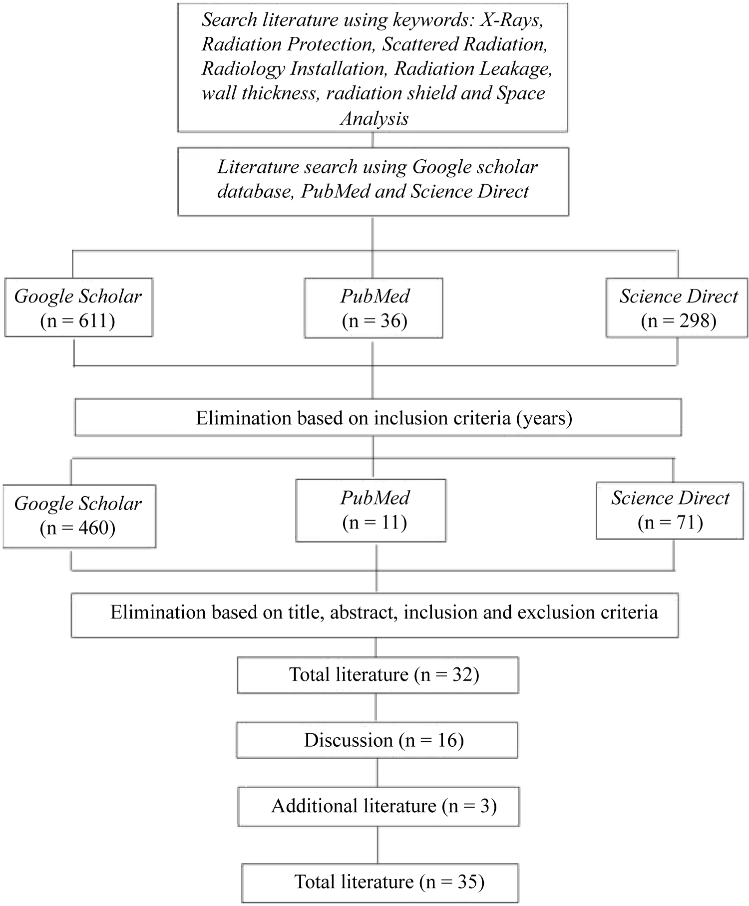 Figure 1: Literature search and selection chart.
View Figure 1
Figure 1: Literature search and selection chart.
View Figure 1
X-rays are widely used in the health sector, one of which is in radiodiagnostics. The use of X-rays must comply with the principles of radiation protection and reception of radiation doses that have been regulated by international standards, namely by the International Atomic Energy Agency (IAEA) and BAPETEN. According to BAPETEN PERKA Number 4 of 2013, radiation workers may not receive radiation doses exceeding 50 mSv per year and the average per year should not be more than 20 mSv, while the general public should not be more than 1 mSv per year [9].
X-ray radiation is not only beneficial in the health sector, it can also have a detrimental effect on society and the surrounding environment if it is excessive. The greater the dose of scattered radiation received, the greater the negative impact of radiation that can be received. Therefore, it is necessary to control the negative effects by paying attention to and applying radiation protection. The basic principles of radiation protection are distance, shielding and time [4]. Several things must be considered before the installation of an X-ray aircraft room, including the location of the building, the location and design of the room, as well as the thickness of the walls and door shields and glass [10].
Based on a number of studies, radiation protection efforts to limit scatter radiation exposure in terms of space analysis can be carried out in various ways as summarized in Table 1.
Table 1: Radiation protection efforts to limit exposure to scattered radiation are viewed from the analysis point of view of the room. View Table 1
Table 1 shows that a number of health facilities carry out radiation protection efforts with various efforts, including setting distance, voltage, height of radiation source, thickness of radiation shield, position of radiation source, and installation of radiation warning signs. The research was carried out using various types of X-ray machines such as CT-Scan, dental panoramic, and several types of X-ray machines which are used for general medical and dental examinations. In addition, several other tools are used, such as a survey meter which is used to measure the radiation dose rate, a meter to measure distance, and a phantom head which is used as a patient substitute. From several research results reviewed, most researchers focus more on setting the distance and voltage of the radiation source.
Based on the above discussion, it can be concluded that there are many protective measures that can be taken to limit exposure to scattered radiation doses. Taking into account the distance and the magnitude of the voltage can have a major effect on the rate of exposure to radiation doses received by officers and the general public in the vicinity of the radiation source room.
One of the radiation protection efforts to reduce the intensity of radiation emitted and reduce the radiation dose received by the human body is the manufacture of radiation shields because radiation shields can absorb some of the radiation. The greater the effectiveness of a room's radiation shield, the better the radiation shield will absorb radiation [11]. Radiation shields are made according to the thickness of the walls of the room that have been determined in the applicable regulations so that people outside the radiation room are safe from the dangers of X-ray radiation [1].
There are several types of materials used and the thicknesses needed to build walls in radiation facilities, including red brick with a thickness of 25 cm and a density of 2.2 g/cm3 or concrete with a thickness of 20 cm or equivalent to 2 mm of lead (Pb), so that the exposure received does not exceed the dose limit value of 1 mSv/year [12]. An explanation of the effect of retaining wall thickness in limiting scatter radiation exposure is shown in Table 2.
Table 2: The thickness of the retaining wall in limiting exposure to scattered radiation. View Table 2
The results of the studies in Table 2 are in accordance with the theory discussed by Sutejo & Daryati [13], which explains that the thicker or denser the radiation shield structure, the less radiation that can pass through the screen. Table 2 also shows that there are different types of materials used in the radiation facilities studied. Types of materials for the manufacture of radiation retaining walls include concrete + lead, plaster + lead, red brick + ceramic. These types of materials have the same effectiveness to withstand scattered radiation exposure doses. To achieve safe radiology room conditions for radiation workers, patients and the public, Bapeten has set a radiation shielding standard, which uses a material equivalent to 2 mm of lead [14].
Lead is a malleable metal that is malleable, shiny in color, shiny after cutting, and soon becomes dull gray when exposed to air. Lead has advantages such as being highly resistant to chemical corrosion and can be used indoors or outdoors, as well as having a high density so that it is very well used as a radiation shield [15,16]. In addition to having advantages, lead has weaknesses that need to be considered, including a high level of toxicity, lead is a toxic substance if absorbed into the body, lowers the development of Intelligence Quotients (IQ), triggers hyperactivity, makes learning difficult, does not care and is aggressive, damage to hearing aids and inhibit growth [14].
Concrete is a mixture of fine and coarse aggregate materials, namely sand, stone, crushed stone, or other similar materials to which cement and water are added. Concrete has advantages and disadvantages. The advantages of concrete include being easy to form, can withstand heavy loads, resistant to high temperatures, low maintenance costs, and easy to obtain, while the weaknesses of concrete are difficult to change the shape when it is formed, require high accuracy, have a heavy weight, as well as great sound reflection [17]. Concrete can be replaced with other alternatives, namely by using lightweight concrete or commonly called hebel. Hebel is made from a dough consisting of quartz sand, cement, lime, a little gypsum, water, and aluminum paste as a developer (chemical air filler). The advantages of Hebel include uniform size and quality, do not require thick casting joints, are lighter than ordinary bricks, construction work is faster than ordinary bricks, does not require thick plastering, waterproof, soundproof, high compressive strength, good resistance to corrosion. Earthquakes, while the disadvantages of Hebel include the need for special adhesives, special skills in installation, relatively more expensive prices than red bricks, and relatively difficult to obtain [16].
From the discussion above, it can be concluded that concrete or lightweight concrete (hebel) coated with lead (Pb) can be used as a radiation shielding material. Concrete with a minimum thickness of 20 cm should be given a lead coating of 2 mm, so that it is able to withstand radiation and does not cause negative effects on the health of patients and radiation workers [2-35].
Effective radiation protection efforts include distance regulation, radiation source voltage and radiation retaining walls. The distance and voltage of the radiation source greatly affect the rate of scattered radiation dose received by patients, radiation workers and the surrounding community. The farther the distance and the smaller the voltage, the smaller the scattered radiation dose rate.
The thickness of the retaining wall affects the rate of scattered radiation dose, the thicker the retaining wall, the smaller the radiation that can penetrate. The selection of the type of retaining wall material can affect the effectiveness of the retaining wall. The type of material that is good for radiation retaining walls is concrete or lightweight concrete (hebel) with a minimum thickness of 20 cm and coated with lead (Pb) with a thickness of 2 mm.
None.
All authors have no potential conflict of interest to declare for this article.