Several measures are applied to characterize the functional state of muscles. Among these, the Maximum Voluntary Contraction (MVC) is most frequently used and considered the gold standard. A little understood and used measure is Neuromuscular Efficiency (NME) of muscles. Therefore, the present study was conducted to compare NME indices of several trunk muscles of healthy adults with respect to age and sex.
Overall 100 healthy Caucasian subjects (equally distributed in young (19-39 years) and elderly (48-71 years) and persons of both sexes, i.e. 25 subjects per age group and sex) were investigated during submaximal and maximal isometric tasks of their trunk muscles in sagittal plane. Five major superficial trunk muscles were evaluated by means of Surface EMG (SEMG). NME and MVC values were extracted. NME was calculated using the inverse slope of the logarithmic transform of the SEMG amplitude to torque relationships of the submaximal tests.
The abdominal muscles showed no sex differences in NME but were always superior in both elderly groups. As could be expected MVC levels during flexion of men were higher but interestingly no age related MVC differences could be detected. However, for the back muscles NME showed no age-related effects and during extension MVC was reduced with age. Males compared with females showed significantly higher NME indices and also higher MVC values. Absolute SEMG amplitudes levels of all investigated trunk muscles were always lower in the elderly subjects.
The NME is an SEMG amplitude independent parameter and may be applied to determine the functional state of (trunk) muscles as a complement to MVC measurements. The observed systematic differences of NME indices between sexes and age groups careful hint towards the NME being related to the functional cross-sectional area of type I fibers.
Surface Electromyography (SEMG), Amplitude-force relationship, Isometric contraction, Maximum Voluntary Contraction (MVC)
ANOVA: Analysis of Variance; BMI: Body Mass Index; CSA: Cross Sectional Area; ECG: Electrocardiogram; EMG: Electromyography; LO: Musculus Longissimus; MF: Musculus Multifidus; MVC: Maximum Voluntary Contraction; NME: Neuromuscular Efficiency; OE: Musculus Obliquus Externus; OI: Musculus Obliquus Internus; RA: Musculus Rectus Abdominis; rms: Root Mean Square; SEMG: Surface Electromyography; SFT: Skin Fold Thickness; UBM: Upper Body Mass; UBT: Upper Body Torque
Adequate physical fitness reduces the risk of musculoskeletal injuries [1]. Therefore, especially in the light of aging populations improvements in diagnosis of neuromuscular performance are of particular interest. Any biomechanically based diagnosis of neuromuscular function considers power (i.e. explosive strength), force, endurance, co-ordination, and efficiency. In addition, Surface EMG (SEMG) can be used to assess major neuromuscular characteristics like endurance, individual strain level, co-ordination, and efficiency and is therefore increasingly used in research and therapeutic settings [2-5].
In research and assessment of the above mentioned neuromuscular characteristics, baseline data need to be established in order to compare subjects or the effect of any kind of intervention such as therapies, training, and nutrition. A simple to take measurement involves determination of Maximum Voluntary Force (MVC). However, an MVC measure can only be an approximation of a subject's force capacity, because it is influenced by motivation [6,7], experience with the required task [8], side dominance [9], tolerance to discomfort and pain [10], age [11], and sex [12,13]. Frequently the MVC is taken as the only measure to assess success of interventions. However, exclusive use of MVC ignores the complexity of neuromuscular function.
An additional way to assess the functional state of a muscle or muscle group is the determination of Neuromuscular Efficiency (NME), a straight forward SEMG application [14,15]. NME has not been extensively investigated and its importance relative to the other mentioned fitness characteristics is still an open question. But, good neuromuscular efficiency simply stands for the achievement of equal force levels with less expense of energy, or equal muscular strain is accompanied by a higher force output [16]. Consideration of NME dates to the early fifties [14-17] and proved to be a predictable measure of the functional state of muscles with respect to sex [18], neurological disorders [19], physical therapy [20], and training [21,22]. Initially, NME indices were suggested to be correlated with morphological changes due to hypertrophy in contrast to neural factors [21,23,24]. More recently it could be shown that NME indices of the rectus abdominis muscle in healthy subjects were higher for physically active in comparison with inactive women and were also higher in males than in females [18].
Initially, NME was calculated as the inverse slope of the linear regression of the EMG to force relationship at submaximal levels [15]. Later on, this initial approach was refined by using logarithmic transformed EMG data [18] to account for the non-linearity of the EMG to force relationship. Due to the calculation algorithm higher NME indices are associated with less steeper slopes of the SEMG amplitude to torque relationship and therefore represent a better efficiency of the neuromuscular system. The NME can also be determined during MVC tasks but is less accurate and in this case it is the ratio between the respective torque and EMG values during maximum effort.
The calculation of NME indices of trunk muscles in the literature is especially rare. One study identified influences of sex and different physical activity levels on the NME of the rectus abdominis muscle [18], another investigation dealt with the alteration of back muscle NME relative to trunk posture [25], and one further study examined the influence of eccentric exercise [22]. As far as we could find studies that simultaneously determined NME of abdominal and back muscles and related these values with maximum force data are completely lacking.
Therefore, the present study was conducted to compare NME indices of main superficial abdominal as well as back muscles with respect to age and sex, and to relate these data to maximum force values to enhance the interpretation of NME indices. These reference data could be used to improve individual diagnostics of a subject's neuromuscular functional state and could therefore enhance prevention, therapy and rehabilitation.
Since the force capacity of females and males differs largely, we expected to find lower NME indices in women compared with men. Further, older subjects were expected to show lower NME indices compared with young subjects, independent of sex.
Overall 100 healthy Caucasian subjects of both sexes, equally distributed in young (19-39 years, mean: 27.3 ± 5.0 years) and elderly subjects (48-71 years, mean 60.1 ± 7.3 years) were included in the study. Detailed information about the study purpose and procedure was provided. Informed consent was obtained from all individual participants included in the study. The study protocol was approved by the ethics committee of the Jena University Hospital (2643-08/09). All participants were clinically examined and asked about their medical history. Exclusion criteria were actual back or radiating pain, any surgery of the spinal column, and also any C-section. Selected anthropometric characteristics are given in Table 1. The level of physical activity on a 1 to 5 self-rating scale was also obtained from all subjects (see Table 1). In this scale 1 stands for "no physical activity at all" and 5 is rated as "extremely high level of physical activity" (daily high intensity sports or hard physical work for at least one hour).
Table 1: Selected anthropometric characteristics and physical activity levels of the study population. Data are given in mean ± SD. View Table 1
All tests were conducted in a computerized test and training device for whole body tilt (CENTAUR, BfMC, Germany). In this device the subject's lower body is fixed at hip and thigh, whereas the upper body remains free (Figure 1). For the submaximal test conditions portions of the Upper Body Mass (UBM) were applied by tilting the subject from vertical position at defined tilt angels. During these tasks the subject had to remain in upright body posture and therefore simply to compensate the gravitational forces acting on the trunk (Figure 1). Correct adherence to upright body position was enabled by a harness, positioned over the subject's shoulder that was equipped with strain gauges. A biofeedback monitor in front of the subject contained a crosshair and a point that was deviated from the center of the crosshair if any force was applied to the harness, i.e. the person failed to maintain the desired upright posture. Exact adherence to the mentioned conditions was controlled by the investigator and assisted by correction suggestions if necessary. The Maximum Voluntary Contraction (MVC) tests were also performed in the device with subjects standing upright.
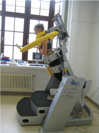 Figure 1: Study setup with a subject positioned in the device, performing a 30° forward tilt. View Figure 1
The applied submaximal test conditions consisted of isometric applications of 9%, 17%, 34%, 50%, 71%, 87%, and 100% of the Upper Body Mass (UBM) and were realized by tilting the subjects by 5°, 10°, 20°, 30°, 45°, 60°, and 90° from vertical position in sagittal plane, i.e. in forward and backward directions. These tilt angles were chosen as a compromise between the possibility of exactly adjustable tilt angles and desired equally spaced torque moment differences. All 14 tasks were applied in an individually randomized order to prevent possible order related effects. While the device moved (i.e. tilting to reach the desired position) subjects leaned relaxed on the harness and were asked to move away from the harness and adopt upright body position if the target angle was reached. Every submaximal task was executed for approximately ten seconds to ensure isometric conditions. The submaximal tests incidentally provided a necessary warmup for obtaining accurate force levels during the MVC test. After completing the submaximal tests, subjects then immediately performed at least one halfhearted practice force test that was followed by three single MVC trials of which the best was used for analysis [26]. For this subject were asked to apply their maximum trunk extension and flexion forces against the harness for approximately three seconds during standing with one-minute interval between each test. Subjects were informed that there would be three MVC trials [26] and strong verbal encouragement was provided [7]. MVC execution was observed religiously and corrected to ensure proper execution. The MVC force data were already registered during the tests. If conspicuous data were observed, i.e. the variability between the tests exceeded more than 10% the MVC test was repeated [27,28].
During all test situations the subjects remained with arms crossed over chest to avoid effects due to varying arm positions.
The UBM of all subjects was determined while the device was tilted forward to 90° and participants lay relaxed on the harness. At this position the strain gauge values corresponded to the subjects' UBM. To ensure plausible values full relaxation of the back muscles was controlled via Surface EMG (SEMG). Hints were provided if remaining activity could be detected. This procedure was repeated two times and the highest value was further used in the analysis. Additionally, the distance between the iliac crest and the projection of the scapular spine to the spine was determined along the spine and further used to calculate the individual upper body torque (UBT, see Table 1) and MVC torque values.
Bipolar SEMG was taken from five superficial trunk muscles, simultaneously from both body sides: M. Rectus Abdominis (RA), M. Obliquus Internus Abdominis (OI), M. Obliquus Externus Abdominis (OE), M. Multifidus Lumbalis (MF), and M. erector spinae, Pars Longissimus (LO). All electrode positions are detailed in Table 2 and were chosen in accordance with the accepted international recommendations and Ng [29-31]. Additionally, the cardiac activity was detected by the application of a further electrode pair along the heart axis for subsequent elimination of the inevitable ECG- contamination of the SEMG signals. Prior to SEMG electrode application the respective regions were marked by an experienced examiner, shaved if necessary, and cleaned with abrasive paste (Epicont, GE, Germany). The used electrodes had a circular uptake area of 1.6 cm diameter (H93SG, Covidien, Germany) and an inter-electrode distance of 2.5 cm. The SEMG signals were amplified (gain: 1000, input impedance: 1200 GΩ, noise level: < 1 µV, CMRR > 120 dB, 10-700 Hz, 1st order RC filter, Biovision, Germany), analog to digital converted (2000/s, Tower of Measurement, 24 bit resolution at ± 5V: 0.6 nV/bit, anti-aliasing filter at 1000 Hz, DeMeTec, Germany), collected (ATISArec, GJB, Germany) and stored on hard drive for offline processing. The MVC force data were determined by simultaneously capturing the strain gauge values together with the SEMG data. To determine the MVC force levels steady sections of the force graph correlating to the three MVC trials were marked. For each of these sections the mean was calculated, and the highest value was used as the respective MVC force level.
Table 2: SEMG Electrode positions. View Table 2
Raw data were band-pass filtered between 10 Hz and 300 Hz. Possible interferences from the power supply were eliminated by a 50 Hz notch filter. To eliminate the inevitable ECG contamination of the SEMG signals only steady-state sections of 400 ms, starting at an interval of 100 ms subsequent to all determined R-waves, were considered for analysis. The average value of all respective segments was used to calculate the representative root mean square (rms) values per trial and muscle. The NME of the abdominal muscles was determined using data from all backward tilt positions (i.e. isomeric trunk flexion tasks) and for the NME of back muscles all the forward tilt data were combined (i.e. isomeric trunk extension tasks).
NME can be calculated from either MVC tasks or a series of submaximal tasks. To determine the NME during MVC tasks the developed torque or force is related to the respective rms value [32,33]. In our investigation we calculated NME by using a series of submaximal tasks, similar to the approach of David and colleagues [18]. This at first required the identification of the best fit for the respective rms to torque relationships. A second order polynomial function resulted in the best fit (coefficient of determination (r2) 0.971 to 0.991; mean: 0.984, SD ± 0.006) for all muscles, in both sexes and age groups. The rms values were then linearized by application of the natural logarithm. From these logarithmic transformed rms data individual linear regression slopes were fitted. The mean NME for every muscle, sex, and age group was determined by averaging the inverse values of the respective individual linear slopes.
At first, a repeated measures ANOVA was performed to detect if any influence of body side on NME had to be considered and it was deemed there is no side influence. Therefore, pooled values from both sides were used for all subsequent analyses.
Prior to the detailed statistical analyses a univariate ANOVA was performed to identify general influences of muscle, sex, and age on NME. Specific tests for group differences of NME indices were performed by the application of Student's t-tests for independent samples. The significance level was set to p < 0.05. All analyses were conducted with IBM SPSS Statistics 21.0 (IBM, NY, USA).
The univariate ANOVA of the NME indices showed significant influences of age (F = 6.849, p = 0.009), sex (F = 89.392, p < 0.001), and muscle (F = 215.395, p < 0.001), with a significant but ordinal interaction between sex and muscle (F = 25.811, p < 0.001). Therefore, muscles were further analyzed individually and separately for sex and age.
The NME indices of the abdominal muscles differed with age, always being higher in the elderly subjects. This could significantly be shown in males for the RA and OI and in females for the OI (Table 3). In contrast, female subjects always showed significantly lower NME indices for the back muscles independent of age.
Table 3: NME indices of the investigated trunk muscles. View Table 3
The relationship between the SEMG amplitudes together with their logarithmic transforms versus the respective torque levels are detailed in Figure 2. In males SEMG amplitudes at almost all submaximal torque levels differed between the two age groups, being always higher in the young group (Figure 3). However, in females significant SEMG amplitude differences could only be observed in the abdominal muscles, again with higher levels in the young group (Figure 3).
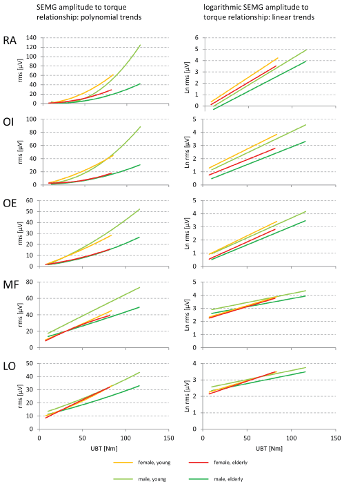 Figure 2: Trend curves for the SEMG to torque relationships of portions of the upper body mass expressed as Upper Body Torque (UBT) values. Left: polynomial trend curves of the respective mean SEMG amplitude to torque relationship, Right: linear trend curves of the logarithmic transforms of the mean SEMG amplitude to torque relationship. View Figure 2
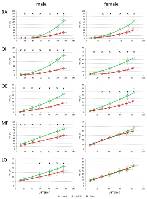 Figure 3: SEMG amplitude to torque relationships of the investigated male (left) and female (right) subjects. The statistics were performed under the assumption of equal UBT values. All data are given as mean ± SD. View Figure 3
For both flexion and extension muscle groups the MVC torque levels were always significantly higher in male subjects. For the maximum extension task young women showed significantly higher torque levels in comparison to the elderly female group (Table 4).
Table 4: MVC torque levels. View Table 4
The actual investigation determined NME indices of trunk muscles at submaximal torque levels that were induced by graded tilts in sagittal plane. For the abdominal muscles NME indices were always higher in the elderly subjects, this was especially pronounced in the male group. Interestingly, for the back muscles no systematic age-related influences of the NME could be proven, but women in comparison with men showed significantly lower NME indices. As expected, men in comparison with women always showed higher MVC levels. Interestingly, the MVC levels during flexion (abdominal muscles) were almost identical between both age groups, but for the maximum extension (back muscles) the young groups both performed better - significantly so for the female subjects.
The NME data of the present study are in good agreement with the results of David, et al. [18] and this substantiates a trustworthy basis for the present investigation. They determined NME indices of the RA and MVC flexion torque levels in female gymnasts, male runners, and normally active controls of both sexes. The young males' NME indices of the RA in the present study are positioned just midway between the NME indices of the male controls and the runners of the David study. The NME indices of our young female group correspond more to the group of gymnasts and not their normal female controls. As to the MVC torque data our female young group's data perfectly match with the Davis young normal controls data, but our young males performed more like the male runners. Because both MVC and NME data of our young males were better than in the David investigation it might be that our young males were somehow fitter. To further support the correspondence of these two studies, the BMI of our young groups of both sexes fit nicely with those of the respective sex groups [18].
If NME is redundant with either MVC and SEMG amplitudes then one would expect concordant differences between age groups and between sexes, but this is not the case: NME, independent of sex, increased with age for all tested muscles for both flexion and extension, while MVC during extension together with all SEMG amplitude levels decreased with age and remained virtually unchanged during flexion. By looking at sex differences we found the NME to be higher in males for the back muscles only. As expected MVC levels, again together with the SEMG amplitude levels, were always higher in males in both flexion and extension exercises.
With increasing age, due to age related involution, there is a loss of alpha and gamma motor neurons [34,35] together with a numerical atrophy that is particularly pronounced in type II fibers [36]. Data from healthy young subjects of both sexes are available detailing fiber type and distribution of thoracic and lumbar back muscles [37]. Mannion and colleagues showed that the cross-sectional area of all muscle types was considerably larger in men [37]. Further, the ratio of type I fibers is much higher in back muscles as compared with the abdominals [38, 39].
Combining these morphological facts helps to hypothetically explain the found differences in NME indices, since they are in concordance with the expected Cross Sectional Area (CSA) of type I fibers according to age, sex, and specific muscle (i.e. localization).
Beside the involution related arguments, the elderly subjects showed an elevated BMI. This was independent of sex. By definition the calculation of the NME is independent from amplitude level. At this point it cannot be excluded that the reduced amplitudes in the elderly may have yet influenced the results, most probably due to irregular dampening conditions for low and high muscular strain levels. This argument also remains hypothetical, since to the authors' knowledge no systematic investigations exist in this regard.
Since we did not measure the fiber-type distribution in our subjects the given hypothetical interpretation of NME still contains a missing link that has to be closed by future investigations.
The present study was conducted using the CENTAUR that applies graded forces on the trunk by tilting the subjects in sagittal plane. This offers the advantage of highly reliable testing, but still is an artificial situation.
A second order polynomial function was the best fit of the amplitude to torque relationship of all investigated trunk muscles. However, the non-transformed SEMG values the back muscles showed a very low quadratic coefficient (mean values MF: -0.00023, LO: 0.00046) that would allow the application of a linear regression with high accuracy [40], but one would not have been able to directly compare NME data between back and abdominal muscles. Therefore, by generally using the logarithmic transformed data any unaware error due to misleadingly applied linear calculations for the non-transformed values of the abdominal muscles can be avoided.
Summarizing all mentioned aspects NME indices might be a correlate of the functional cross sectional area of type I fibers: The number of type I fibers is always higher in the back muscles and the CSA of all fiber types is larger in men. Further, with increasing age primarily the number of type II fibers is reduced. This affects the abdominal muscles first since they contain a larger proportion of type II fibers.
The NME seems to be a force independent but morpho-functionally linked characteristic of muscle function that is independent from SEMG amplitude level and maximum force (MVC). It may therefore be applied as a physiologically derivable indicator of the type I to type II ratio, but this is currently speculative. Based on the known age-related change in fiber type, it may be reasonable to infer that the elderly showed higher NME indices of their abdominal muscles and men showed larger NME indices of back muscles than women.
SEMG amplitudes are influenced by many factors. Because of this it is recommended to normalize SEMG amplitudes. To account for interindividual differences in this study we measured the BMI and the SFT. Neither one of these was able to explain all the observed differences. This is in agreement to previous theoretical [41,42] and practical [43] results: Accounting for subcutaneous fat failed to sufficiently explain interindividual amplitude variance of SEMG amplitudes [43,44].
The NME of trunk muscles shows differences according to sex, age, and specific muscle. The calculated NME indices are robust against both systematic SEMG amplitude differences and MVC levels. The NME therefore offers a promising complementary, physiologically based measure to assess the functional state of trunk muscles that may indirectly hint at fiber type distribution, although at this point this is mostly speculative. The NME could then be used to specifically guide and monitor rehabilitation and training. In as far as how well a certain protocol is supporting the increase of the one or the other type of muscle fiber (type I for increased strength and type II for increased stability) the NME would then change or not. Anyhow, this hypothesis needs to be proven by specific training interventions.
The authors wish to thank all the subjects for their time and effort. Measurements were carried out at the kindly provided laboratory of the Center for Interdisciplinary Prevention of Diseases related to Professional Activities (KIP) funded by the Friedrich-Schiller-University Jena and the German Social Accident Insurance Institution for the foodstuffs and catering industry. The authors also gratefully acknowledge the linguistic and intellectual support by Ms. Marcie Matthews of polished words.
The study was supported by the Central Innovation Program of the German Federal Ministry of Economics and Technology, Grant KF2150501WD8. The study sponsors had no involvement in the study design, the collection, analysis and interpretation of data, or in the writing of the manuscript. Hereby all authors decline any financial and personal relationships with other people or organizations.